


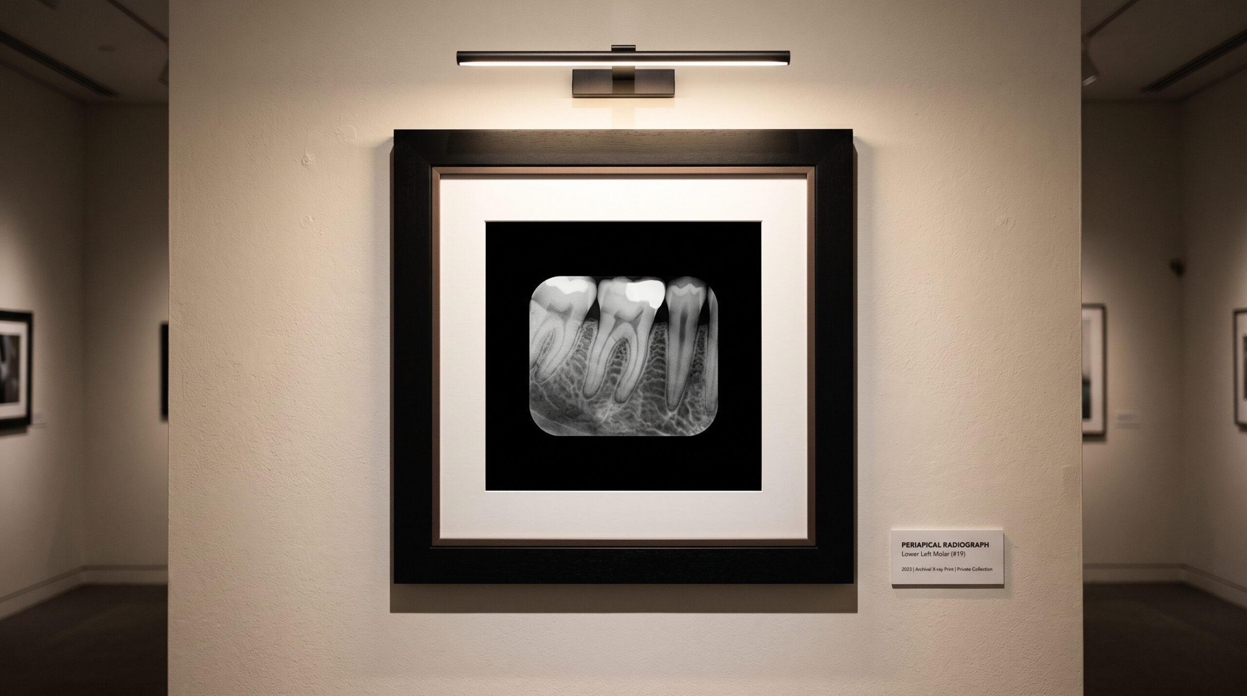
February 8, 2026 The Photon-Counting Detector: A New Physics Beneath the Radiograph
Hold a conventional dental radiograph up to the light and you are looking at an average. Across the fraction of a second the beam was on, a scintillator glowed, a photodiode gathered that glow into a well of charge, and the electronics read the total. Bright where many photons landed, dark where few did. It is an elegant compromise, and it has served dentistry faithfully for a century. But it is, fundamentally, a summing — and everything that summing discards, we have simply learned to live without.
The photon-counting detector refuses to sum. It counts each X-ray photon as a discrete event, one at a time, and it decides — photon by photon — whether that event is worth recording at all. This is not a better version of the sensor we already have. It is a different physics beneath the image, and the difference is legible in the picture.
Two Ways to Turn an X-Ray Into a Picture
Every digital dental sensor faces the same first problem: an X-ray photon is invisible to a photodiode. Something must translate it. The overwhelming majority of intraoral systems solve this indirectly. A scintillator layer — cesium iodide or gadolinium oxysulfide — absorbs the X-ray and re-emits its energy as visible light, which a CMOS or CCD array then reads. In a 2025 survey of intraoral systems in Imaging Science in Dentistry, fully 95.2% used this indirect approach; 80% of those were CMOS.
The scintillator is also where sharpness goes to die. Light scatters sideways as it travels to the photodiode, spreading a single photon’s signal across neighbouring pixels — a soft, luminous bleed that no amount of downstream processing fully recovers. Direct-conversion sensors delete the middleman. A semiconductor — historically amorphous selenium, now increasingly cadmium telluride (CdTe) — absorbs the X-ray and generates electrical charge directly, with no visible-light detour and far less lateral spread. In that same survey, direct conversion accounted for under 5% of systems. Photon-counting lives inside that thin slice of the market — which is precisely why it is worth understanding now, before it is everywhere.
Counting, Not Integrating
Here is the pivot. A conventional detector is energy-integrating: it lets charge accumulate for the whole exposure and reads the pile at the end. Every source of noise that accumulates alongside the signal — dark current, leakage, read noise — is baked into that pile. It is the faint grey haze that sits under every radiograph, the reason a subtle lesion at the edge of the greyscale is a judgement call rather than a fact.
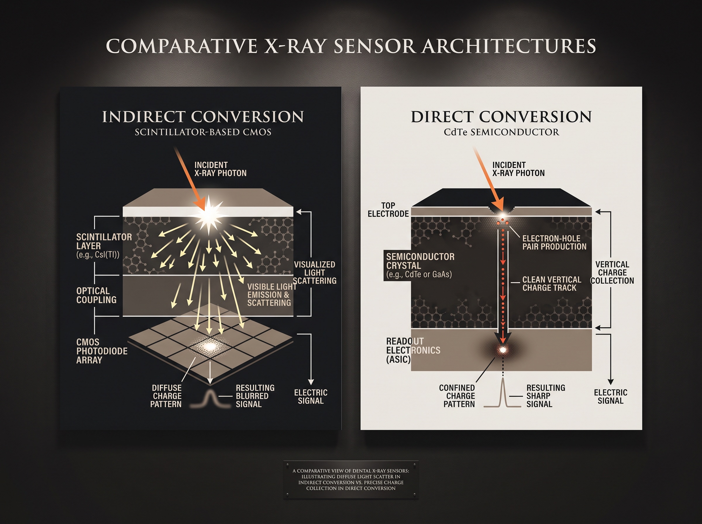
A photon-counting detector instead watches the semiconductor in real time and registers a count every time an arriving photon deposits more than a set energy threshold. Set that threshold just above the electronic noise floor, and the noise is not subtracted — it is never counted in the first place. The detector reports zero dark current. The greyscale no longer floats on a haze; it begins at true black. For an imaging craft that lives and dies on low-contrast detectability — the incipient caries, the hairline vertical root fracture — starting from true black is not a cosmetic gain. It is diagnostic headroom.
The Material-Science Bargain: Charge Sharing
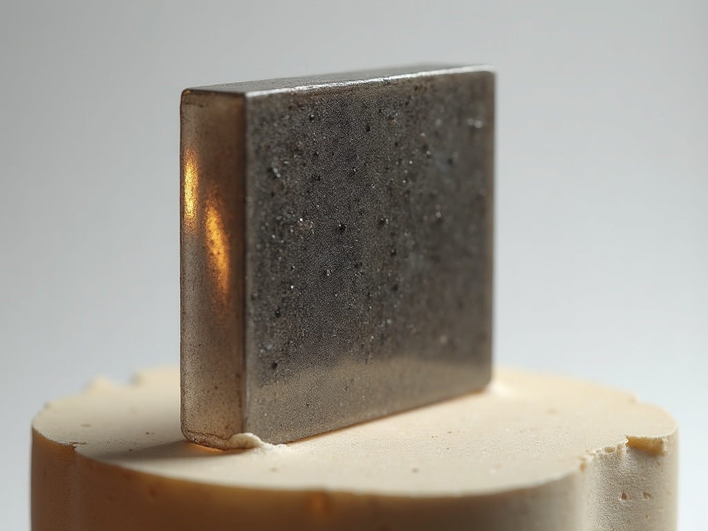
None of this is free, and the price is paid in materials engineering. CdTe is chosen because its high atomic number makes it a greedy absorber of diagnostic X-rays in a thin layer — the panoramic detector described in a 2023 Biomedical Physics & Engineering Express study used a CdTe sensor just 0.75 mm thick. But when a photon lands near a pixel boundary, the cloud of charge it liberates spills across two or three pixels. Left uncorrected, one photon gets counted several times, at fractions of its true energy — smearing the image and corrupting the very energy measurement that makes counting worthwhile.
The answer is charge-sharing correction: on-chip logic that recognises a split event, sums the fragments, and assigns the photon to a single pixel at its full energy. The same 2023 detector ran two adjustable energy thresholds with charge-sharing correction at up to 300 frames per second. That a correction of this subtlety now happens silently, in hardware, at video rates, is the quiet triumph that makes clinical photon-counting plausible at all.
Colour in a Grey World: Spectral Imaging
Once you are measuring the energy of every photon, a radiograph stops being purely grey. Set two energy thresholds and you can bin photons into a low-energy and a high-energy image from a single exposure — and because different materials attenuate high and low energies differently, you can begin to tell them apart. Enamel from dentine. Bone from a metallic artefact. A restorative material from the tooth it sits against.
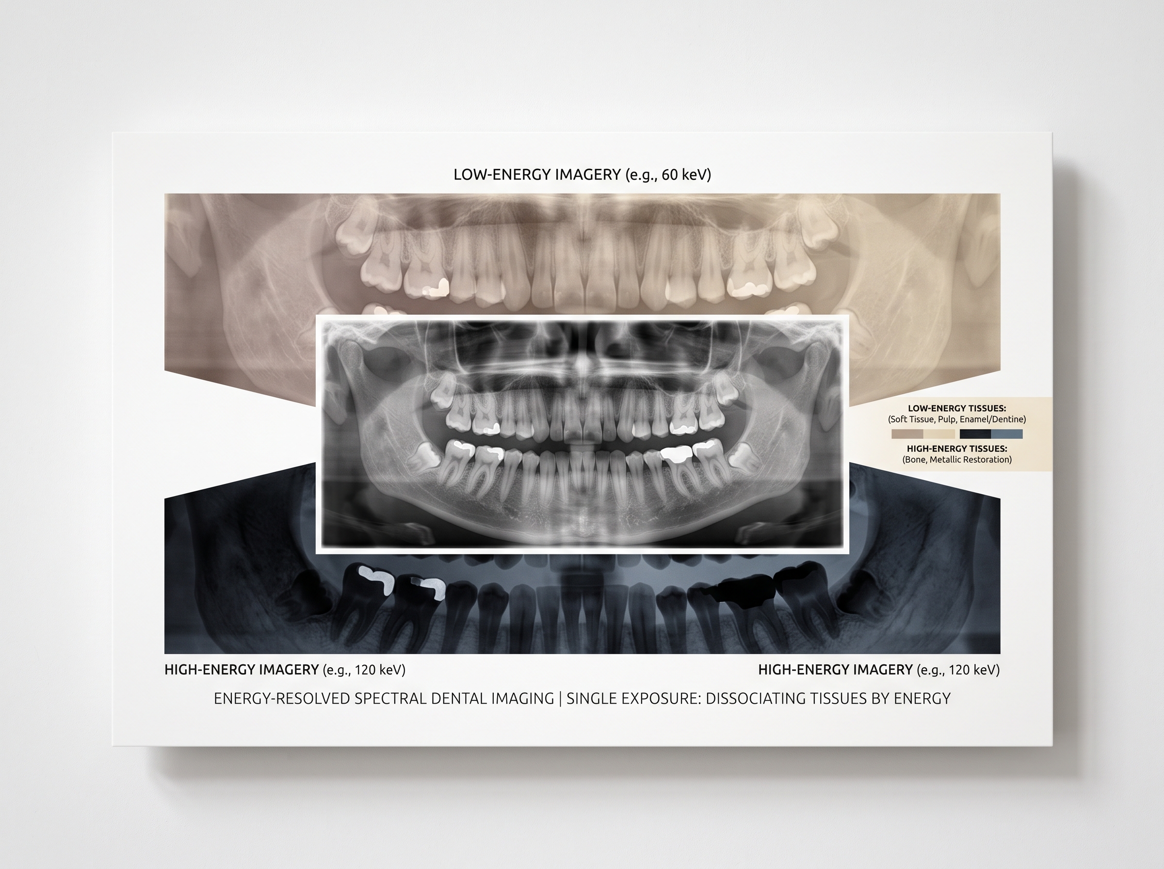
This is spectral — or material-discriminating — imaging, and it is the capability energy-integrating detectors physically cannot offer, because they threw the energy information away the moment they summed the charge. In dentistry it remains early: the panoramic work above is a research platform, not a shipping product. But the door it opens — a radiograph that encodes composition, not merely density — is the kind of door that does not close again.
From Laboratory to Operatory
The temptation is to file all of this under “someday.” That would be a mistake. Direct-conversion intraoral sensors have already crossed into commerce — DentalTI markets its DC-Air as the first intraoral sensor to convert X-ray photons directly to image data, promising a cleaner image and thinner profile without a scintillator’s blur. The full photon-counting, energy-discriminating intraoral sensor is not yet a catalogue item, but the components — thick CdTe, fast per-pixel counting electronics, on-chip charge-sharing correction — have all shipped in adjacent products.
What this means for the clinician who cares about the image is a coming realignment of expectations. Lower dose for equal information, because no photons are wasted feeding a noise floor. Higher effective resolution, because the light-scatter penalty of the scintillator is gone. And, eventually, a radiograph that answers questions of what a structure is, not only where it sits.
Future Developments
The through-line of dental imaging’s last two decades has been the steady removal of intermediaries between the photon and the picture — film to phosphor plate, plate to scintillator-and-CMOS, and now scintillator to bare semiconductor. The photon-counting detector is the logical terminus of that arc: nothing stands between the X-ray and its measurement but the atoms chosen to absorb it. As CdTe growth matures and per-pixel electronics shrink to intraoral scale, expect the first genuinely spectral intraoral sensors to appear as premium instruments, then to migrate downward as every new modality eventually does. When they arrive, the radiograph will have quietly changed its nature — from an averaged shadow into a counted, characterised record of the tissue it depicts. The image, as ever here, is both the science and the art; the photon-counting detector simply gives us more of the truth to render beautifully.
Sources & further reading:
- Intraoral digital radiography: technical specifications of current and historical systems (Imaging Science in Dentistry, 2025)
- Spectral photon counting for panoramic dental imaging (Biomedical Physics & Engineering Express, IOPscience)
- DC-Air Wireless Direct-Conversion Intraoral Sensor (DentalTI)
Related Reading
- Detective Quantum Efficiency: The True Measure of a Dental Sensor, Beyond Resolution
- Quantitative Light-Induced Fluorescence (QLF) Imaging: Revolutionary Caries Detection Technology in Modern Dentistry
- Radiopaque Contrast Agents in Dental Imaging: Advanced Material Science for Enhanced Diagnostic Visibility
Sorry, the comment form is closed at this time.