


August 25, 2026 The Shadow of Protection: What a Bismuth Thyroid Shield Costs a Panoramic Radiograph
There is a gesture every dental team knows by heart: the soft collar laid gently around a patient’s neck before the exposure. It reads as unambiguous care – a small shield placed over a vulnerable gland, a promise that the practice takes protection seriously. And yet, in one of the quiet ironies of imaging, this most tender gesture is also the one most likely to ruin the very picture it accompanies. A thyroid shield is an act of protection that, on a panoramic radiograph, tends to photograph as a wound.
A recent study in Scientific Reports examined a flexible thyroid shield built from a bismuth-oxide silicone-rubber composite, measured against a Rando phantom, and its findings sit precisely on the fault line the profession has been reckoning with: the shield works, and that is not the whole question. To understand why protection and image are so often at odds here, you have to look at what bismuth actually does to a beam, and at the peculiar way a panoramic machine builds its picture.
What Bismuth Does to a Beam
Bismuth is a heavy element, and heaviness is the entire point. With a high atomic number, its dense nucleus is an efficient absorber of diagnostic-energy X-rays through the photoelectric effect – photons are swallowed rather than passed, and the dose that would have reached the tissue behind it is genuinely reduced. Formed into a silicone composite, it becomes a lead-free, drapeable collar that is lighter and more comfortable than traditional lead, and the phantom dosimetry confirms it does lower the exposure delivered to the thyroid.
But absorption is never the only thing a dense material does. The same interactions that stop primary photons also generate scattered radiation, and a high-Z shield placed in or near the beam becomes a secondary source of stray, low-quality photons. Those scattered rays do not carry clean anatomical information; they wash into the image as fog and interfere with the primary beam that does. This is the same family of physics that governs how scatter and collimation shape contrast in a radiograph – except here the scatter is being manufactured by the protective device itself, at the worst possible place.
The Tyranny of Rotational Geometry
An intraoral film forgives a collar because the source is a single fixed cone; place the shield below the field of view and it never appears. Panoramic imaging grants no such mercy, because it does not take a picture so much as sweep one. The X-ray source and detector orbit the patient in a slow arc, building the final image strip by strip as the beam travels through the sharply defined layer we call the focal trough.
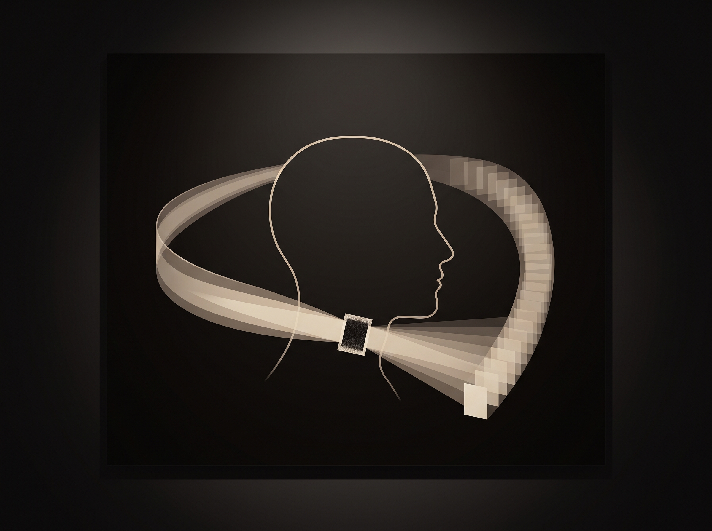
A shield that sits still while the source moves is therefore not photographed once – it is swept through, its dense edge intercepting the beam at a continuously changing angle. The result is the signature failure: a broad radiopaque band or ghost rising across the lower border of the image, most often obscuring the anterior mandible, symphysis, and the roots of the lower incisors. It is the same rotational logic that produces the ghost shadows a panoramic machine paints where no bone exists, turned against a protective device. The anatomy hidden behind that band is not merely dimmed; it is gone, and no window or level adjustment can recover information the shield never let reach the sensor.
An Artifact Has a Dose of Its Own
It is tempting to file this under aesthetics – an ugly image, but a safe patient. That framing misses the real cost. When a shield obscures diagnostic anatomy, the honest response is a repeat exposure, and a retake means the entire panoramic dose is delivered a second time, this time with the collar removed. The gesture meant to spare the thyroid a small dose has instead doubled the exposure to every other structure the beam passes through. Protection that provokes a retake is not protection; it is dose laundering.
There is a subtler cost, too. A band across the mandible does not always announce itself as an artifact – it can be quietly read as pathology, or it can simply hide a lesion that was there. An image degraded at the point of care carries a diagnostic risk that outlives the appointment, in the same way that unaccounted exposure quietly accumulates when a practice stops watching the numbers, as it does with exposure index and dose creep. The artifact is not free. It is paid for later, by someone.
When the Guidelines Changed Their Mind
For decades the thyroid collar was treated as an unqualified good, and questioning it felt like questioning safety itself. The evidence has since forced a reappraisal. Modern dental imaging – rectangular collimation, fast digital receptors, tightly limited fields – delivers effective doses so low that the incremental risk to the thyroid from a panoramic or cephalometric exposure is, in the assessment of the American Dental Association’s expert panel, negligible. On that basis the guidance now recommends that thyroid shielding not be used routinely for intraoral, panoramic, cephalometric, or cone-beam imaging.
This is not a dismissal of protection; it is the same disciplined weighing that governs the choice between radiation risk and diagnostic risk in cone-beam imaging. When the dose spared is vanishingly small and the probability of a shield-induced artifact – and its accompanying retake – is real, the arithmetic inverts. The collar that once symbolized caution becomes, in the panoramic context, a net source of dose and a reliable spoiler of images. The refined position is not to shield harder, but to collimate precisely, image only what is indicated, and let the machine’s own restraint do the protecting.
Future Developments
The bismuth-composite study is valuable precisely because it refuses the easy answer. It confirms that better materials can lower dose while shedding the weight of lead, and that line of work matters for the settings – certain CBCT fields, pediatric protocols, anxious patients who need the reassurance – where a shield may still earn its place. The frontier is a collar engineered around the geometry rather than against it: shaped, positioned, and bounded so its edge never enters the focal trough’s sweep, pairing genuine attenuation with an image left untouched. Materials science is quietly dissolving the old trade-off between the dose we can stop and the picture we must preserve. Until that shield exists, the most sophisticated protection a panoramic exposure can offer is often an honest, well-collimated beam and the discipline to leave the neck unclothed – trusting the numbers, and refusing to cast a shadow across the very anatomy we came to read.
Sources & further reading:
- Shahbazi L, Sardari D, Malekie S, et al. Evaluation of a flexible thyroid shield in dental panoramic radiography utilizing bismuth oxide silicone rubber composite and Rando phantom. Scientific Reports 16, 541 (2026)
- American Dental Association Council on Scientific Affairs. Patient shielding during dentomaxillofacial radiography. JADA (2023)
No Comments