


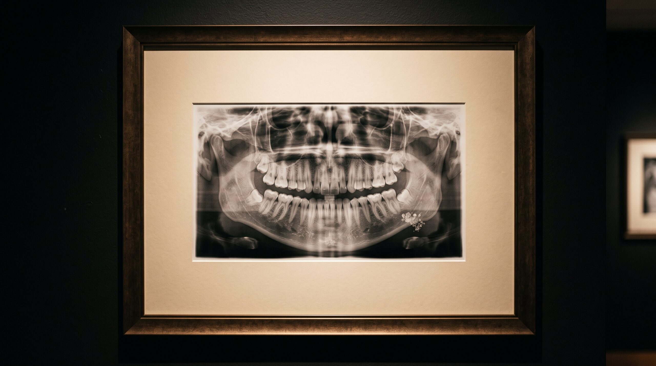
November 12, 2026 The Panorama as Sentinel: Carotid Calcifications and the Radiograph That Reads the Whole Body
Every image is ordered for a reason, and the reason narrows the eye. A panoramic radiograph is requested to survey the dentition, the jaws, the sinuses, the condyles; the clinician’s attention travels the arch from one ramus to the other and stops where the teeth stop. Yet the panoramic machine does not know what it was asked to find. It sweeps a tomographic slice through the whole lower face and neck, and in that generous frame it captures tissues that have nothing to do with teeth: the hyoid, the airway, the cervical vertebrae, and the soft corridor of the neck where the great vessels run. Sometimes, in that corridor, the image returns something unbidden — a faint cluster of radiopaque flecks, like popcorn scattered below the angle of the jaw. It is easy to sweep past. It may also be the first visible warning of a diseased carotid artery.
The Stipple Below the Jaw
A calcified carotid atheroma has a characteristic radiographic signature, and learning to see it is a matter of knowing where to look and what to expect. The findings sit inferior and posterior to the angle of the mandible, in the soft tissue adjacent to the third and fourth cervical vertebrae, roughly at the level of the hyoid. They present as small nodular or vertically oriented radiopacities — a stippled or popcorn texture rather than a single dense mass — and they are frequently bilateral. The location is the whole tell. These marks lie well below and behind everything the radiograph was commissioned to show, in a region the reading eye crosses only if it has been trained to travel there deliberately. The discipline is not unlike the one required to separate normal anatomy from true pathology elsewhere on the film: it begins with knowing that the margins of the image carry information too.
What the Mineral Is Telling You
Calcification is the body’s slow record of injury. In the carotid, an atherosclerotic plaque forms at the bifurcation of the common carotid artery — a site of turbulent flow where the vessel divides into its internal and external branches — and over years the plaque mineralises. That mineral is what the radiograph catches. The presence of a calcified plaque does not, by itself, grade the degree of stenosis, nor does it forecast a stroke; a heavily calcified plaque can be stable, and a dangerous one can be soft and invisible to film. What the finding does carry is association. Carotid calcification correlates with the broader burden of atherosclerotic disease and with the familiar roster of risk — hypertension, diabetes, chronic kidney disease, smoking, dyslipidaemia. It is a flag planted in the imaging record, and its meaning is epidemiological before it is individual.
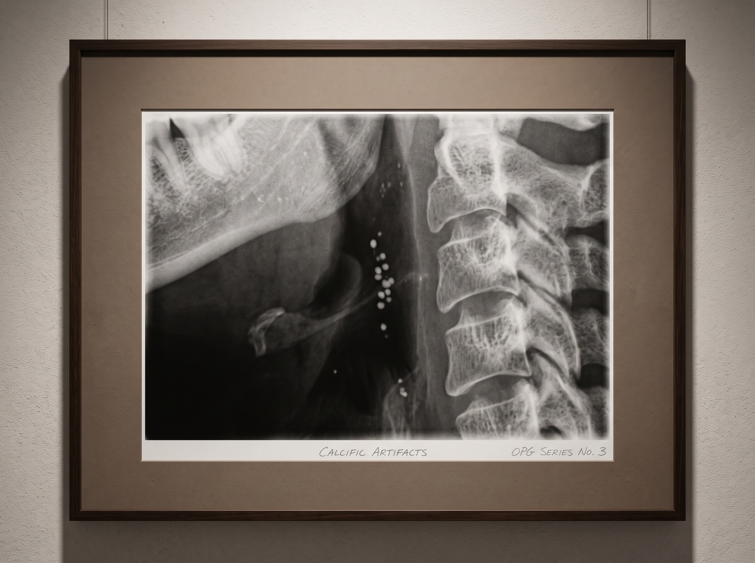
This is also why the finding demands humility in its reading. A dentist does not diagnose vascular disease from a panoramic film, and should never imply otherwise to a patient. The soft-tissue opacity could represent a calcified lymph node, a phlebolith, a tonsillolith, or calcified cartilage of the larynx — each with its own location and morphology. Distinguishing them is the interpretive work, and when the pattern and position point to the carotid, the clinician’s role is narrow and clear: recognise, document, and refer. The image opens a question; the physician and a duplex ultrasound answer it.
The Panorama Is Not the Only Sentinel
The carotid is the most discussed example, but it is one instance of a larger truth about imaging. A radiograph taken for one purpose almost always contains evidence of another. A chest film ordered for a cough carries the shadow of the aorta and the heart. A lateral spine image for back pain frames the abdominal aorta, where calcification predicts cardiovascular events. Even a routine bitewing or periapical, narrow as its field is, occasionally frames a tonsillolith or a styloid process of startling length. The panoramic simply happens to be dentistry’s widest regular window onto the neck, and so it is dentistry’s most frequent accidental messenger. Reading it well means accepting that the frame is always larger than the referral — a theme that runs through how the panoramic geometry itself projects structures into unexpected places.
When the Machine Learns to Watch the Margins
Here the story turns toward the near future, because the one weakness of opportunistic findings is that they depend on a tired human eye travelling to the edge of the image at the end of a long day. Calcifications are missed not because they are invisible but because they are peripheral to the task at hand. This is precisely the gap that machine vision is built to close. Through 2026, deep-learning models have grown unexpectedly good at reading systemic risk from images taken for other reasons — estimating cardiovascular risk from a plain chest radiograph with an accuracy rivalling established clinical scores, and flagging aortic calcification on spine films acquired for orthopaedic complaints. The models do not tire, and they do not have a referral to narrow their attention.
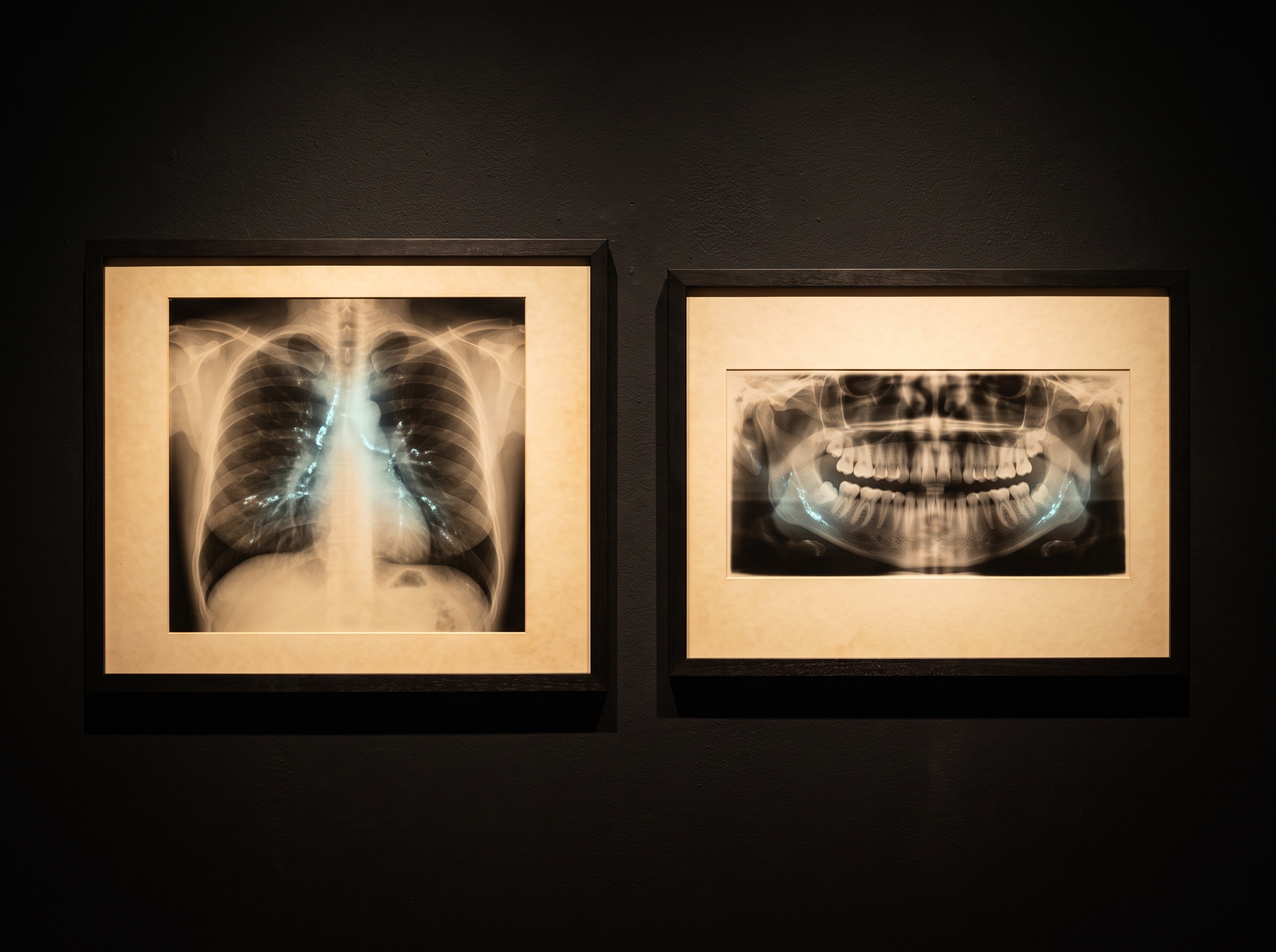
The implication for dental imaging is direct. A model trained to detect soft-tissue calcification on panoramic radiographs would turn the carotid finding from a lucky human catch into a consistent, reproducible prompt — a quiet flag appended to the report whenever the stipple appears below the jaw. The value is not that the machine diagnoses; it is that the machine never fails to look. The same architectures now being used to read caries and bone loss across imaging modalities can be pointed at the neck, and the output can be made legible rather than opaque, so the clinician sees why the model highlighted a region and can confirm or dismiss it with judgement intact. The best version of this technology does not replace the reading eye; it extends the eye’s reach to the parts of the frame it was never asked to inspect.
The Ethics of Looking Further
Opportunistic screening carries a quiet ethical weight. Once an image can reveal more than it was ordered to show, the question of responsibility follows. A clinician who recognises a probable carotid calcification and says nothing has failed the patient; a clinician who over-reads every neck opacity into alarm has failed them differently. The right posture is calibrated attention: a trained habit of glancing deliberately at the cervical soft tissues on every panoramic, a measured threshold for what warrants mention, and a standing pathway for referral that treats the finding as a beginning rather than a verdict. As automated flags enter the workflow, this calibration becomes a shared duty between the algorithm’s sensitivity and the clinician’s restraint. The image has always held more than we asked of it; the obligation is simply to look honestly at what it offers.
Future Developments
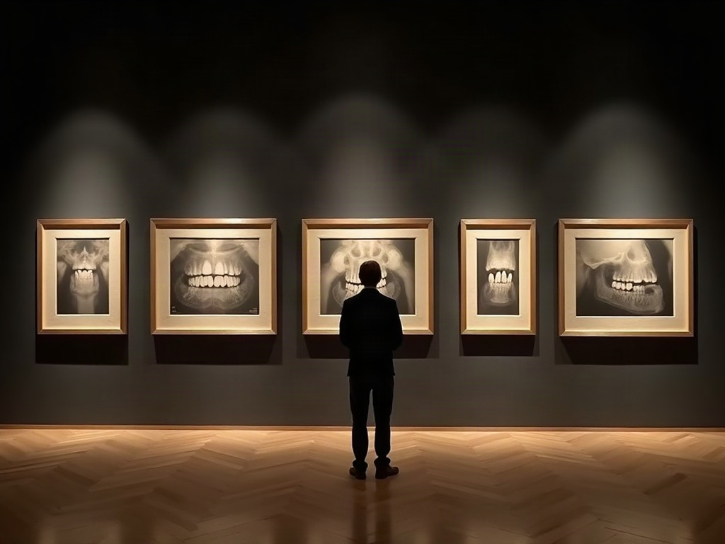
The panoramic radiograph was conceived as a map of the jaws, and for a century that is how it has been read. What is changing is not the image but our relationship to its margins. As models learn to watch the quiet corridor of the neck, as opportunistic screening moves from anecdote to protocol, and as explainable outputs let clinicians trust what the machine marks, the dental radiograph quietly earns a second vocation — not as a cardiovascular diagnostic, but as a patient, ever-present sentinel that happens to see the whole body in the course of surveying the mouth. The art of imaging has always been the art of seeing what is there rather than only what was sought. In the faint popcorn stipple below the angle of the jaw, the next chapter of that art is already visible, waiting for an eye — human or trained — to travel to the edge of the frame and read it.
Sources & further reading:
- Deep learning on chest radiographs predicting cardiovascular risk (research coverage, 2026)
- ADA/FDA guidance on dental radiographic examinations and interpretation
No Comments