


October 24, 2026 The Jaw That Moves on the Screen: Fusing a Still CBCT With Live Motion Tracking
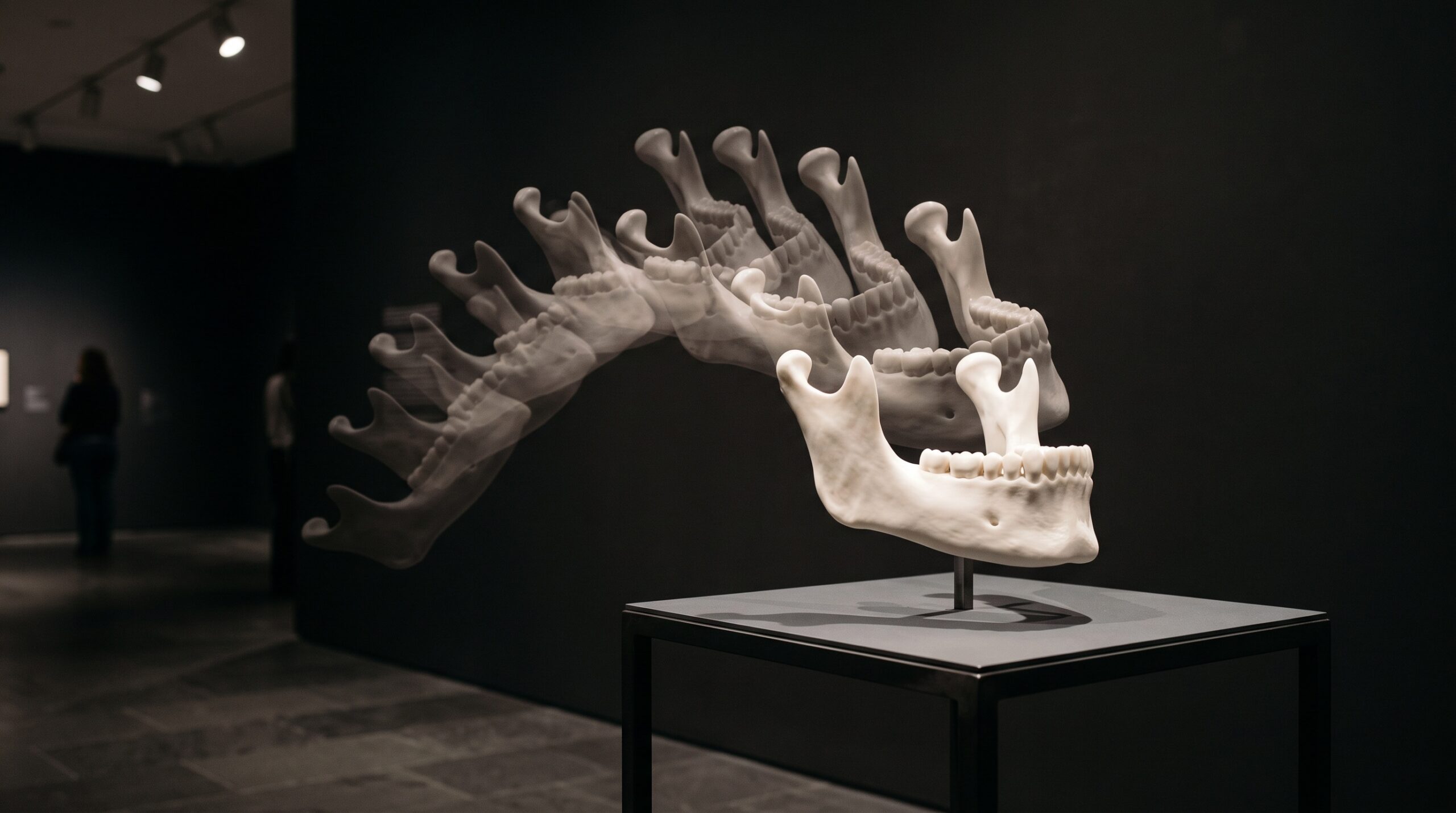
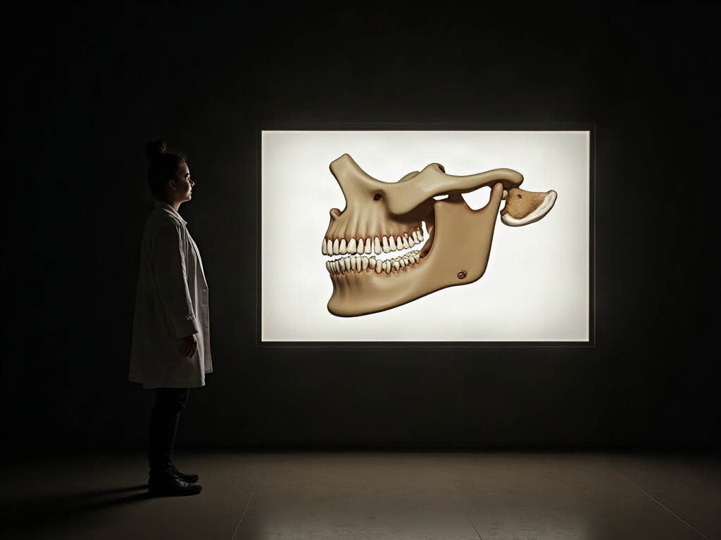
There is a quiet irony at the heart of three-dimensional dental imaging. A cone-beam scan gives us the jaw in extraordinary stillness — every condyle, cortex, and cusp tip held in perfect, frozen register, a volume we can rotate and slice at will. And yet the mandible is the most restlessly mobile joint complex in the body, a hinge that also slides, a structure whose whole clinical meaning is bound up in how it moves. The still scan, for all its exquisite detail, captures the one state in which the jaw almost never finds itself: motionless. For decades we accepted that trade, reading function by inference from a single arrested instant. A maturing class of imaging refuses the trade. It takes the still volume and marries it to a live, high-rate recording of movement, so that the anatomy we scanned can be watched performing. The result is not a photograph of the jaw but a performance of it — and it asks us, as a gallery of the image always should, to look again at what we thought we had already seen.
Two recordings, one coordinate frame
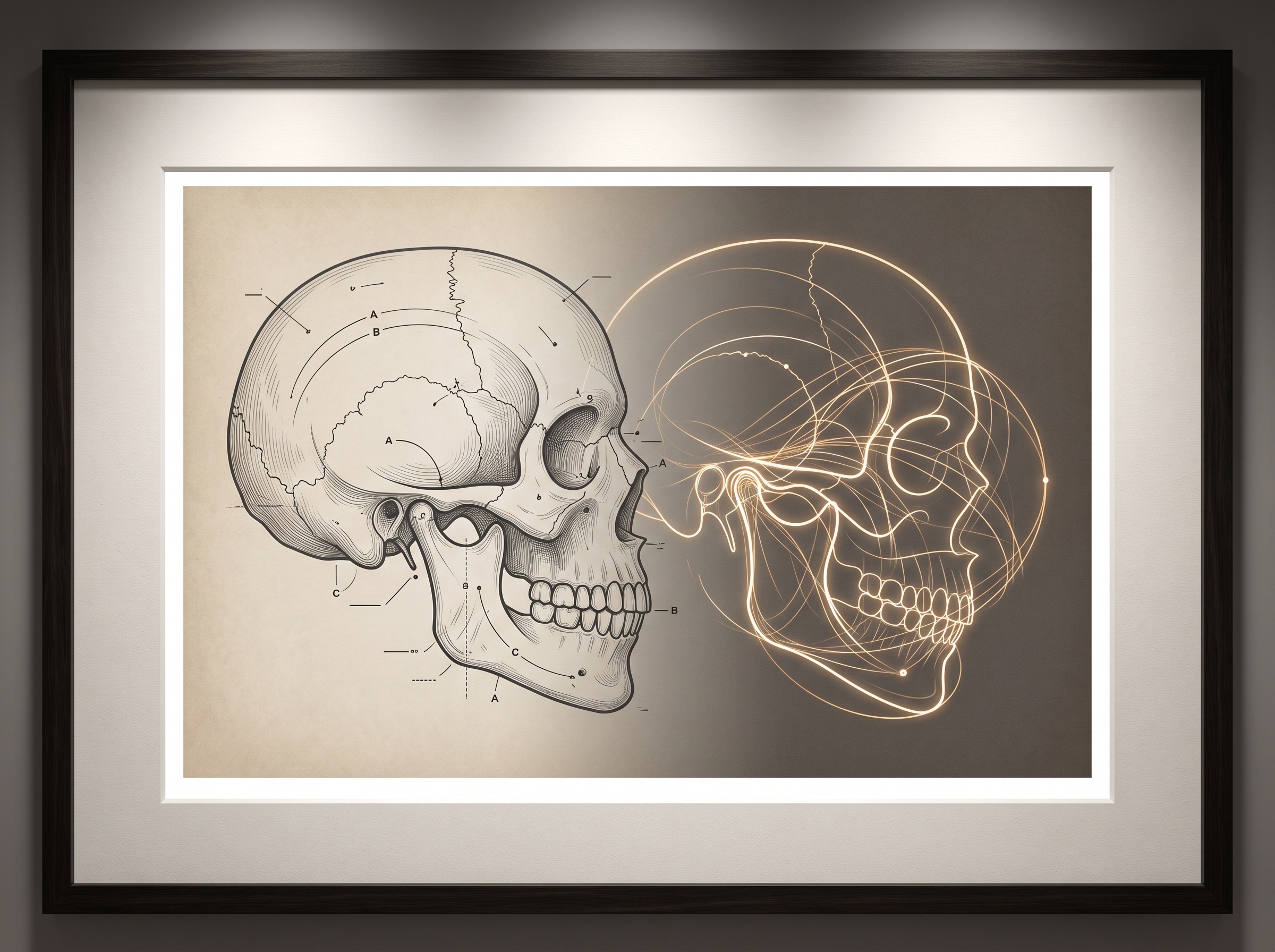
The idea is deceptively simple and the execution is anything but. On one side sits the static anatomy: the CBCT volume, often fused with an intraoral scan of the dentition and sometimes a facial scan, assembled into the composite we have described before as the virtual patient built from face scans, CBCT, and intraoral data. On the other side sits movement: a jaw-tracking device records the position and orientation of the mandible many times a second as the patient opens, closes, protrudes, and chews. The magic — and the engineering difficulty — lies entirely in registration: in placing those recorded trajectories into the same coordinate frame as the scanned skeleton, so that a point tracked in the room corresponds exactly to a point in the volume. Get the registration right and the still condyle in the scan begins to travel along the real path the patient’s own condyle traced. A 2026 workflow published in the prosthodontic literature laid out exactly this marriage, integrating static anatomical registration with a six-degree-of-freedom recording of jaw motion to build a functional virtual patient. Six degrees of freedom is the crucial phrase: the mandible does not merely swing open on a fixed axis but rotates and translates along all three axes at once, and only a recording that captures all six can tell the truth about how a given jaw moves.
Why the hinge refuses to be a hinge
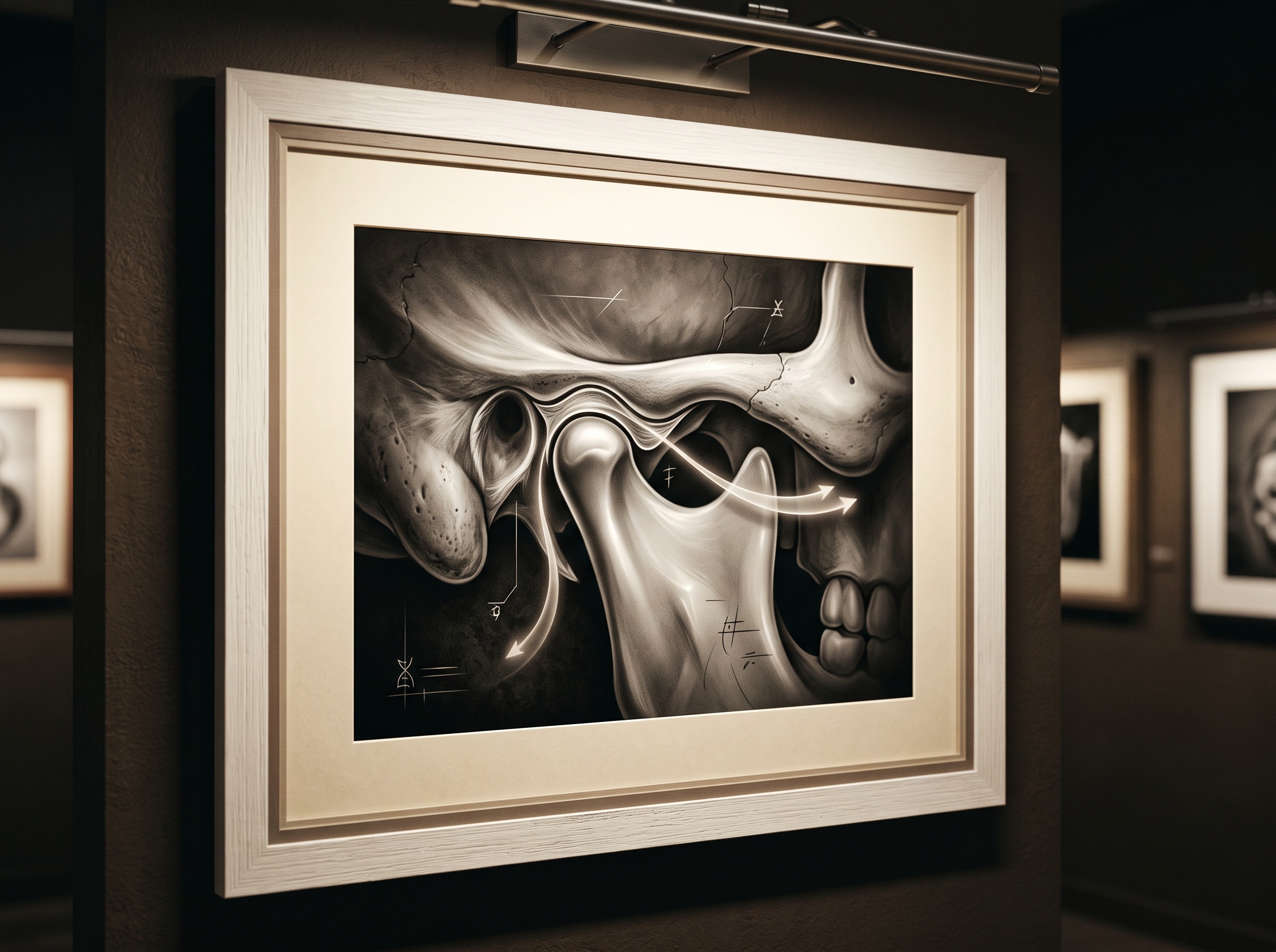
The temporomandibular joint we have learned to picture with MRI and CBCT is the reason none of this can be faked by a mechanical stand-in. A traditional dental articulator approximates jaw movement with average anatomical values — a generic condylar inclination, a textbook Bennett angle — and for many cases that average is close enough. But the real condyle seated in its fossa does not rotate like a door hinge; it rolls and slides down and forward along the articular eminence, on a curved path that is as individual to a patient as a gait. When a restoration, a full-arch rehabilitation, or an orthognathic plan has to respect that path, substituting the average for the actual is precisely where occlusal interferences and premature contacts are born. Capturing the true condylar trajectory is what devices such as the optical Modjaw system and the ultrasonic Zebris were built to do, and the literature on them is consistent on the point that matters: they record the patient’s genuine hinge-axis kinematics during functional movement, not a programmed approximation of it. Fused back onto the CBCT, that trajectory turns the static joint into a joint we can watch work.
From a volume to a dynamic twin
What this fusion produces is best thought of as a dynamic digital twin of the masticatory system. In software, the scanned dentition can be driven through the patient’s own recorded movements, and the clinician can watch — frame by frame, from any angle — where the teeth actually touch, slide, and separate across an excursion. This is a different order of information from the static bite we have always verified by eye and articulating paper, or even from the elegant three-dimensional checks of vertical dimension we explored in our look at stereophotogrammetry and the proof of a restored bite. A static check confirms where the arches meet at one chosen position; the dynamic twin shows the entire choreography between positions — the working and balancing contacts, the guidance that disoccludes the back teeth as the jaw moves forward, the interference that only appears three millimetres into a lateral excursion. It extends the same impulse that drives four-dimensional capture of the moving smile inward from the face to the joint: both insist that a feature defined by motion cannot be honestly recorded by a single still frame.
The honest ledger
None of this is free of friction, and a gallery that respects the image owes the reader the caveats. The hardest of them is registration cost. Some optical systems — the Planmeca 4D Jaw Motion platform among them — have historically relied on a second CBCT exposure with physical trackers fixed to the arches, simply to locate those trackers precisely within the volume; that is additional radiation spent not on anatomy but on bookkeeping. Newer approaches push hard against this: the Modjaw and SDIMatrix systems accommodate tracker repositioning through digital model registration and so avoid the extra scan, and research groups are piloting infrared jaw-tracking with modified bite-fork registration precisely to pin movement onto the scan without that second dose. There is also the perennial question of accuracy — soft-tissue flex at the tracker mount, the sub-millimetre error budget of the registration, the fidelity of the chewing motion a patient performs while wired to an instrument in an unfamiliar clinic. These are not reasons to withhold the technology; they are the terms on which it is honestly used. The fused four-dimensional record is a model, and like every model it is only as trustworthy as the registration that built it.
Future Developments
The trajectory here points toward motion capture that leaves no footprint of its own — markerless jaw tracking that reads the mandible’s movement from ordinary optical or depth sensors and registers it to the scan by the shape of the teeth alone, retiring both the fixed trackers and the second CBCT. Couple that with reconstruction and segmentation already learning to label the volume automatically, and the dynamic twin could assemble itself from a single low-dose scan and a few seconds of natural chewing, ready to drive a restoration design or a splint through the patient’s real envelope of motion. There is something fitting in where this leaves a gallery devoted to the still image. We spent a century learning to hold the jaw perfectly still so we could see it clearly, and the next advance is to let it move again — to understand that for this most mobile of structures, the truest portrait was never the one that asked it to stop. The frozen scan showed us the instrument. Only the moving picture lets us hear it played.
Sources & further reading:
- Adaptive digital workflow for virtual patient modeling: Integrating static registration and six-degree-of-freedom jaw tracking
- A pilot study on an infrared jaw-tracking method for mandibular motion based on modified bite-fork registration: Accuracy and feasibility
- Virtual Dental Articulation Using Computed Tomography Data and Motion Tracking
No Comments