


November 16, 2026 Eagle in the Panorama: The Incidental Findings a Dental Pan Catches Beyond the Teeth
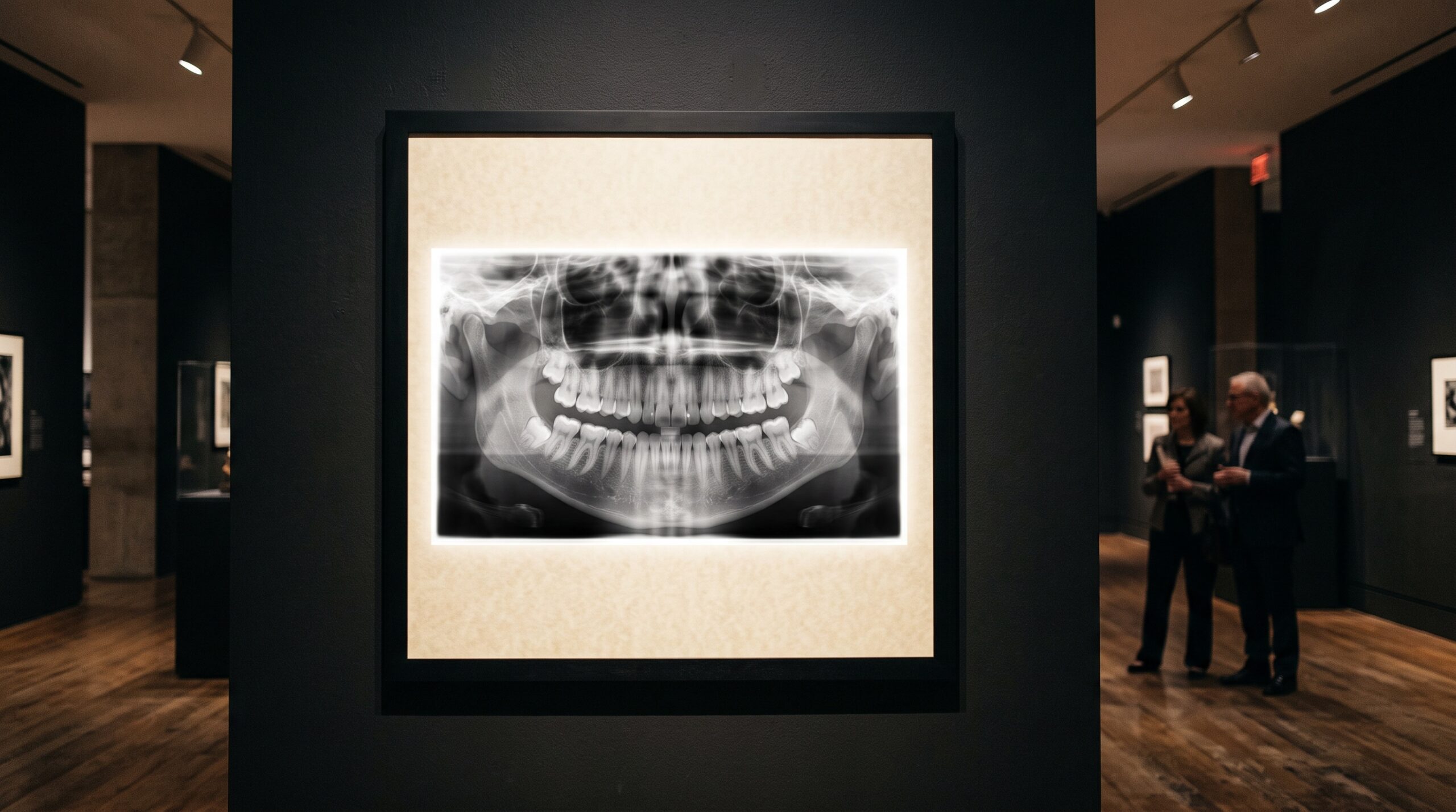
A panoramic radiograph is ordered for the teeth. It is requested to settle a question about third molars, to survey bone before an implant, to confirm the stage of a developing dentition. Yet the image it returns is far larger than that question. The panoramic beam sweeps an arc from one condyle to the other, and in doing so it records the maxillary sinuses, the nasal floor, the orbital rims, the hyoid, the styloid processes, the soft-tissue outline of the airway and the upper reach of the cervical spine. The teeth are simply the subject the clinician came for. The rest of the frame is a quiet exhibition of everything else the beam happened to pass through.
To treat that frame as if it contained only teeth is to hang a large canvas and look at one corner. The craft of panoramic interpretation — the part that separates a glance from a reading — is the deliberate refusal to stop at the dentition. What follows is a short tour of the gallery beyond the teeth, and of the discipline required to actually see it.
The Eagle in the Image: the elongated styloid process
Descending from the temporal bone, just in front of and below the ear, is a slender bony spike called the styloid process. On a well-positioned panoramic radiograph it often appears as a thin radiopaque needle pointing down and forward across the mandibular ramus toward the hyoid. In many people it is short and unremarkable. In others it is strikingly long, or the stylohyoid ligament suspended from it has ossified, and the result is a prominent spur that reads almost like a second, misplaced bone laid over the neck.
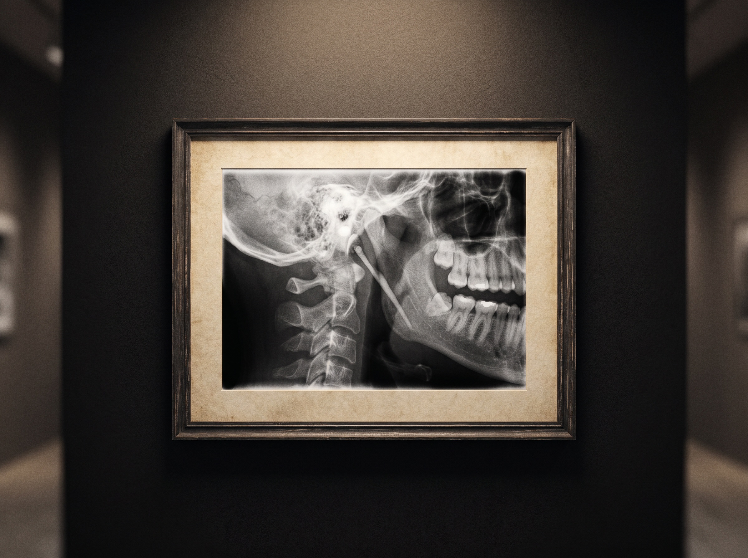
An elongated styloid process is, on its own, a radiographic observation rather than a diagnosis. When it is accompanied by symptoms — a nagging sore throat, pain on swallowing, a catch when the head is turned, a sensation of something foreign in the throat — the constellation is named Eagle syndrome. Most elongated styloid processes never announce themselves; they are found incidentally on an image taken for an entirely dental reason. But the radiographic appearance is characteristic enough that it deserves to be named in the report and, when symptoms line up, correlated clinically. The panorama is frequently the first place the finding is ever seen.
Stones in the soft tissue: tonsilloliths and calcifications
Superimposed over the middle of the ramus, where the airway shadow crosses the bone, small rounded radiopacities sometimes cluster in a loose group. These are often tonsilloliths — calcified concretions within the tonsillar crypts — projected onto the mandible by the geometry of the panoramic sweep. They are usually harmless, but they are a reminder that the panorama flattens a three-dimensional head into a single plane, so a shadow sitting “on” the bone may in fact lie well behind it in the soft tissue of the throat.
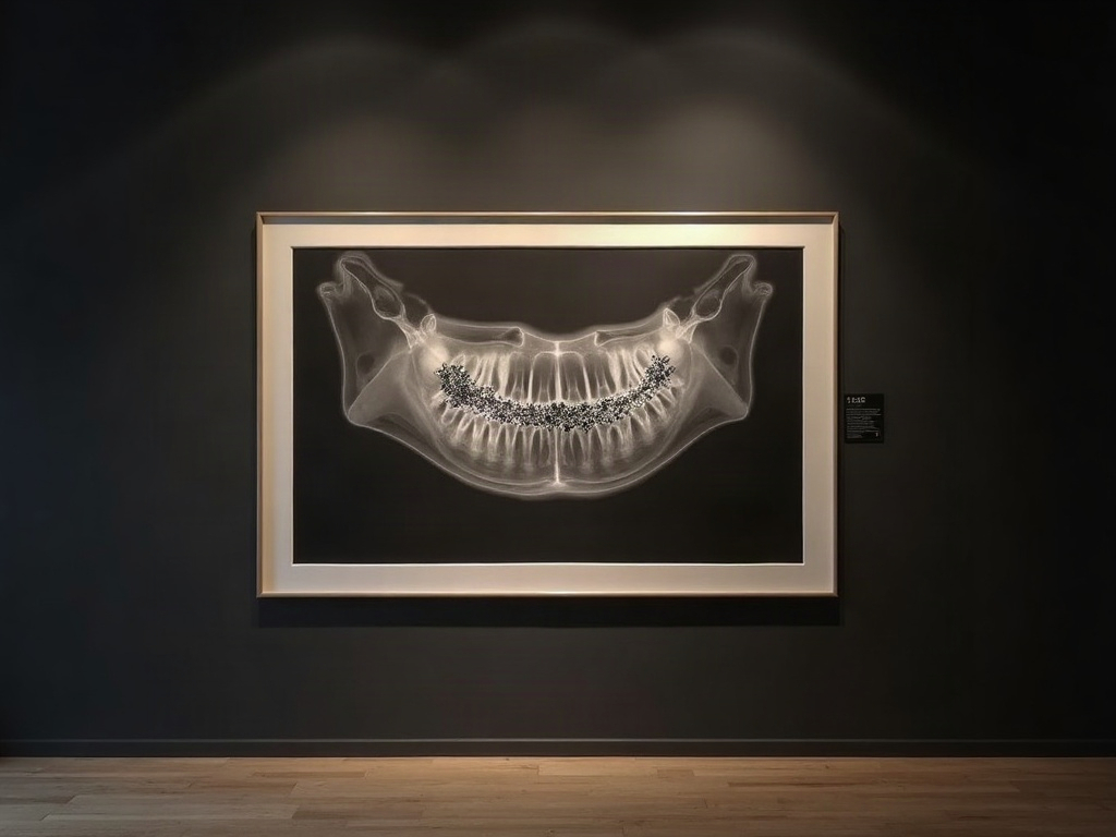
The same plane captures other calcifications: in the submandibular region a radiopacity may represent a sialolith in the gland or duct; lower and more posterior, rounded or laminated opacities near the hyoid demand the careful distinction discussed below. Recognizing that a calcification is soft-tissue rather than osseous is half the interpretation, and it is a skill in reading superimposition as much as in recognizing the object itself. It is the radiographic cousin of the problem we have written about in distinguishing normal anatomy from pathology on the dental image — a shadow that looks like disease, or disease that hides as a shadow.
The sinus overhead: mucosal thickening and pseudocysts
The floors and lower walls of the maxillary sinuses sit directly above the posterior maxillary teeth, which is why the panorama captures them so readily. A gently thickened mucosal lining, a smooth dome-shaped opacity rising from the sinus floor — a retention pseudocyst — or a more diffuse clouding are all common incidental sights. Most are benign and asymptomatic, but they matter for a practical reason: the posterior maxilla is exactly where sinus-adjacent extractions and implant planning happen, and the state of the sinus changes the conversation.
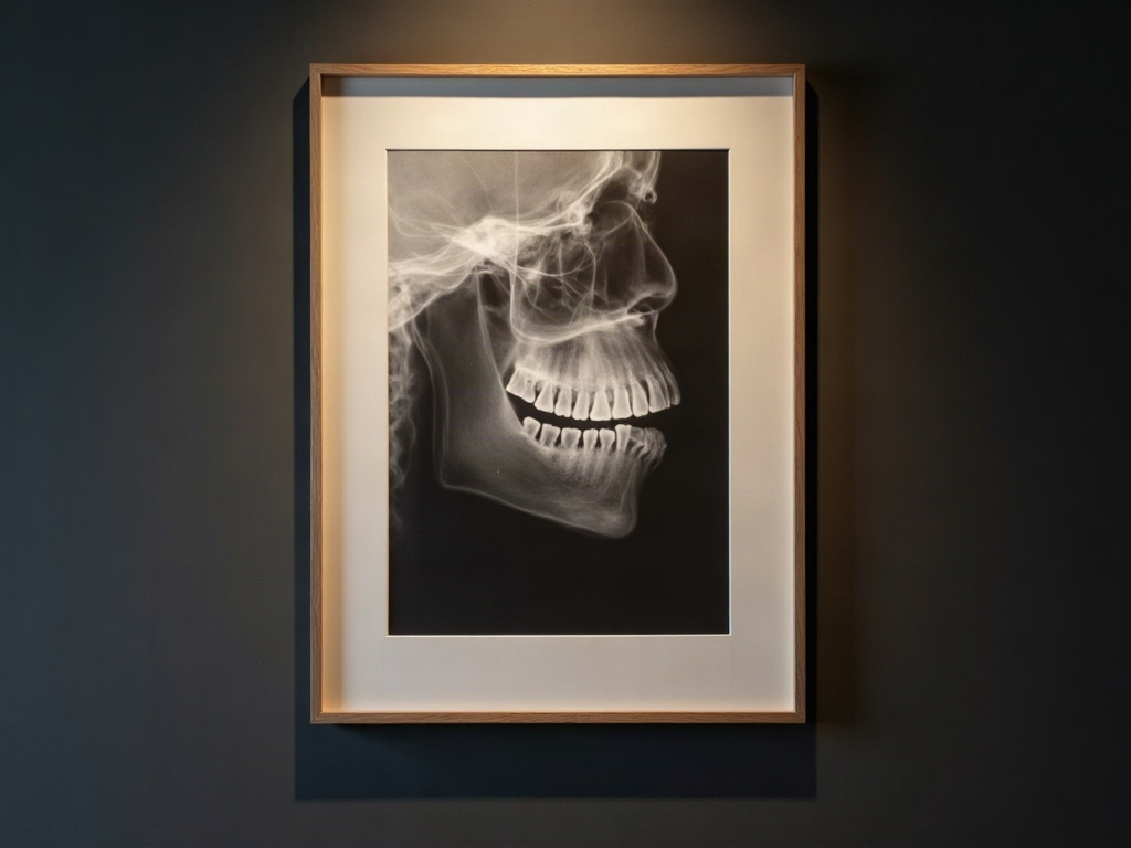
Here the panorama shows its limits as honestly as its reach. A two-dimensional projection can suggest sinus involvement but cannot define it, and this is one of the clearest cases where a cross-sectional study earns its dose. That the same clinical question is answered very differently by a flat image and a volumetric one is a theme in its own right, one we explored in how interpretation shifts across intraoral, panoramic and CBCT imaging.
The airway and the shadows that breathe
Between the hard structures, the panorama also records air and soft tissue: the dark column of the oropharyngeal airway, the dorsum of the tongue, the soft palate, sometimes the shadow of enlarged adenoidal or tonsillar tissue in a younger patient. None of this is diagnostic of a sleep or breathing disorder, and the panorama is not the instrument to make that call. But a markedly narrowed airway shadow or conspicuous lymphoid tissue is a legitimate prompt — a reason to ask a question, to note the observation, to point a colleague toward the right test. The image did not set out to study breathing; it merely declines to hide it.
Down the neck: the cervical spine and the carotid question
At the lower edge of many panoramic images, the cervical vertebrae and the region of the carotid bifurcation drift into view. Degenerative changes at the vertebral bodies can be visible, and near the level of C3–C4 two findings must be told apart: the benign, often laminated triticeous cartilage calcification sitting within the laryngeal cartilages, and a carotid artery calcification, which carries very different weight. Mistaking one for the other, in either direction, is a real interpretive trap.
The carotid finding deserves — and has — its own full treatment, because a calcified atheroma glimpsed on a dental image can be the first hint of systemic vascular disease; we devoted a separate piece to the panorama as a sentinel for carotid calcifications. The point to carry here is narrower: the lower corners of the panorama are not throwaway margins. They are where the dental image quietly touches general medicine, and they reward the same careful looking as the teeth at the center.
The discipline of the whole-image read
Every finding above shares one enemy: satisfaction of search. Once the eye finds what it came for — the impacted molar, the periapical lesion — it tends to relax, and the periphery of the image goes unread. The countermeasure is not talent but routine: a fixed, repeatable path across the entire frame, condyle to condyle, sinus to hyoid, the same sweep every time, so that the margins are examined by habit rather than by luck.
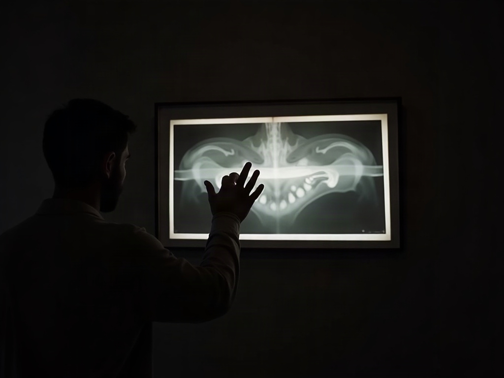
That routine is also where the ethical and medico-legal weight sits. Interpretation is understood to cover the whole captured image, not merely the region of clinical interest. A clinically significant finding that is visible but unrecorded is the uncomfortable scenario behind much of the guidance on radiographic reporting, and the remedy is unglamorous: see it, document it, and refer it when it warrants attention. The panorama is generous with what it shows; the reader’s job is to be equally disciplined in looking, and then to turn a finding into a sentence the record and the patient can both understand — the same translation we described in making the radiograph legible to the person it depicts.
Future Developments
The next chapter of this story is being written by software that does not suffer from satisfaction of search. Detection models trained on whole panoramic images are beginning to flag the very findings a tired eye skates past — the elongated styloid, the sinus opacity, the suspicious calcification near the carotid bulb — not to replace the clinician’s judgment but to insist that the periphery gets a second look. In that collaboration the panorama may finally be read the way it was always capable of being read: as a single image of the lower face, where the teeth are the headline and the rest of the frame is no longer the part we forget. Imaging has always been both science and art; teaching ourselves — and our instruments — to see the whole canvas is how the gallery earns its name.
Sources & further reading:
- American Academy of Oral and Maxillofacial Radiology (clinical standards for radiographic interpretation)
- Incidental findings on panoramic radiographs (medical imaging literature overview)
No Comments