


May 4, 2026 Polarized Light Microscopy in Enamel Analysis: Advanced Birefringence Techniques for Caries Detection and Remineralization Studies
Polarized light microscopy (PLM) stands as one of the most sophisticated and revealing imaging techniques in modern dental diagnostics, offering unprecedented insights into enamel structure and pathology. This advanced optical method leverages the birefringent properties of dental enamel to visualize microscopic changes that are invisible to conventional imaging techniques, making it an invaluable tool for caries detection, remineralization studies, and enamel integrity assessment.
The technique has been employed in dental research for over a century, yet recent advances in digital imaging and analysis software have transformed PLM from a purely observational tool into a quantitative diagnostic powerhouse. By examining how polarized light interacts with enamel crystals, researchers and clinicians can now detect the earliest stages of demineralization, monitor treatment efficacy, and understand the complex dynamics of enamel repair processes.
Understanding Polarized Light Microscopy Principles
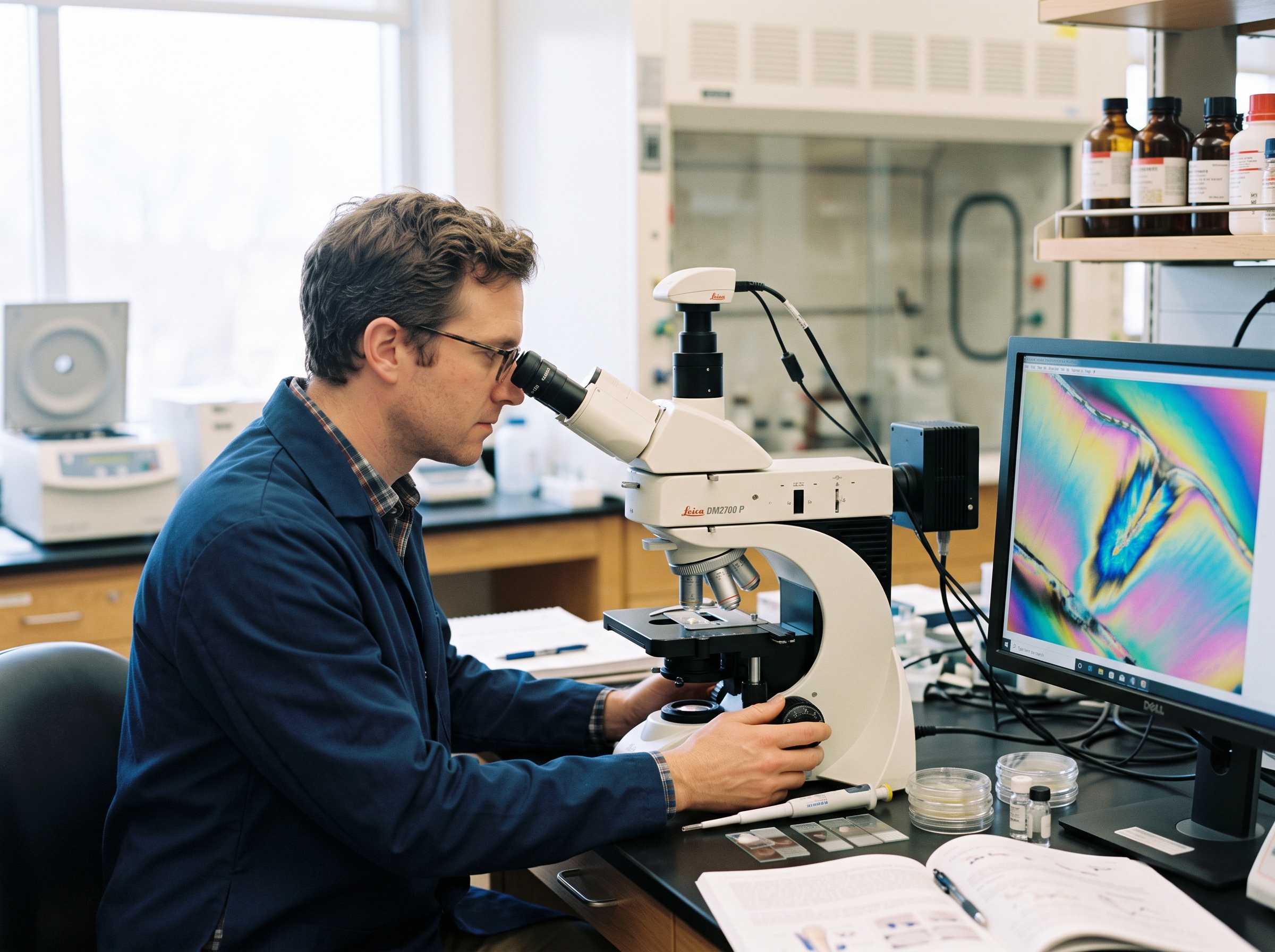
Polarized light microscopy operates on the fundamental principle that dental enamel exhibits birefringent properties due to its highly ordered hydroxyapatite crystal structure. When polarized light passes through enamel tissue, it interacts with these crystals in predictable ways, creating characteristic interference patterns that reveal information about crystal orientation, density, and integrity.

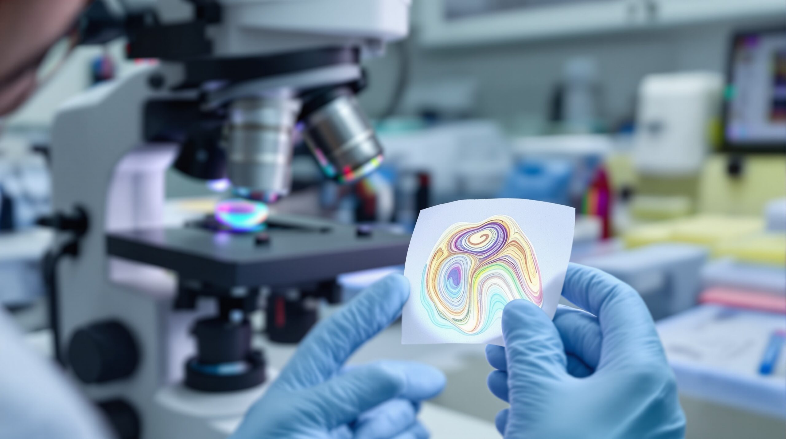
The birefringent nature of enamel becomes particularly evident when specimens are examined under crossed polarizers. Sound enamel typically appears dark or exhibits weak birefringence when immersed in water, while demineralized areas show strong positive birefringence with characteristic rainbow-colored interference patterns. This optical phenomenon occurs because demineralization disrupts the regular crystal structure, altering how light waves interact with the tissue.
Modern PLM systems employ various immersion media, including air, water, and specialized mounting solutions like quinoline, each providing different contrast levels and revealing specific aspects of enamel pathology. The choice of immersion medium significantly affects image interpretation, with quinoline often providing the highest contrast for detecting subtle changes in enamel structure.
Advanced Techniques in Enamel Birefringence Analysis
Contemporary enamel analysis using polarized light microscopy incorporates several sophisticated approaches that enhance diagnostic accuracy. Quantitative polarized light microscopy (qPLM) represents a significant advancement, allowing researchers to measure birefringence values precisely and correlate them with mineral density changes. This quantitative approach transforms subjective visual assessments into objective, reproducible measurements.
Near-infrared polarized light microscopy has emerged as a particularly powerful variant, exploiting the high transparency of dental hard tissues at wavelengths around 1310nm. This approach provides enhanced contrast and deeper tissue penetration, enabling the visualization of subsurface lesions that might be missed by conventional visible-light PLM.
Digital image analysis software has revolutionized PLM applications in dentistry, enabling automated measurement of lesion depth, area calculations, and statistical analysis of birefringence patterns. Modern systems can quantify integrated mineral loss, track remineralization progress over time, and generate detailed reports that support evidence-based treatment decisions.
Clinical Applications and Research Insights
The clinical applications of polarized light microscopy in dental imaging extend far beyond basic caries detection. This technique excels in monitoring the efficacy of remineralizing treatments, providing visual and quantitative evidence of mineral recovery in treated enamel. Fluoride treatment studies, for instance, rely heavily on PLM to demonstrate treatment success by showing the restoration of normal birefringence patterns.
Orthodontic research has embraced PLM for evaluating white spot lesions around brackets, tracking their development, and assessing prevention strategies. The technique’s ability to detect early demineralization makes it ideal for preventive intervention studies and for evaluating the effectiveness of various protective measures.
Quality assurance in dental materials testing also benefits from PLM analysis. The technique can assess the interface between restorative materials and tooth structure, evaluate the effectiveness of bonding agents, and monitor the long-term stability of dental restorations. This application is particularly valuable in developing new materials and treatment protocols.
Advantages and Limitations
Polarized light microscopy offers several distinct advantages in dental imaging applications. The technique provides excellent sensitivity for detecting early enamel changes, requires no special sample preparation beyond sectioning, and offers both qualitative and quantitative analysis capabilities. The non-destructive nature of the optical examination allows for repeated assessments of the same specimen, making it ideal for longitudinal studies.
However, PLM also has limitations that must be considered in clinical applications. The technique requires histological sectioning, limiting its use to extracted teeth or biopsy specimens. Interpretation requires specialized training and experience, and results can be influenced by section thickness, specimen preparation quality, and the choice of immersion medium. Additionally, PLM provides information about mineral density changes but cannot distinguish between different types of bacterial involvement or metabolic processes.
Future Directions and Technological Integration
The integration of polarized light microscopy with other advanced imaging techniques represents the future of comprehensive dental diagnostics. Combined approaches using PLM alongside quantitative light-induced fluorescence, micro-CT imaging, and optical coherence tomography provide multifaceted views of enamel pathology, offering clinicians a complete picture of tissue health.
Artificial intelligence and machine learning applications are beginning to enhance PLM analysis, with automated pattern recognition systems showing promise for standardizing interpretations and reducing observer variability. These technologies could eventually enable real-time analysis and decision support in clinical settings.
Polarized light microscopy continues to evolve as an essential tool in dental imaging, providing unparalleled insights into enamel structure and pathology. Its combination of high sensitivity, quantitative capabilities, and research versatility ensures its continued relevance in advancing our understanding of dental health and disease. As technology continues to advance, PLM will undoubtedly remain at the forefront of innovative dental diagnostic techniques, supporting both clinical practice and scientific research in the pursuit of optimal oral health outcomes.
Related Reading
- Quantitative Light-Induced Fluorescence (QLF) Imaging: Revolutionary Caries Detection Technology in Modern Dentistry
- DIAGNOdent Laser Fluorescence Imaging: Precision Caries Detection Technology for Modern Dental Practice
- Near-Infrared Transillumination (NIRT): Advanced Dental Imaging for Early Caries Detection
Sorry, the comment form is closed at this time.