


November 7, 2026 Where the Premolar Bitewing Really Goes: Tab Placement, Distal Canine Coverage, and Killing the Overlap
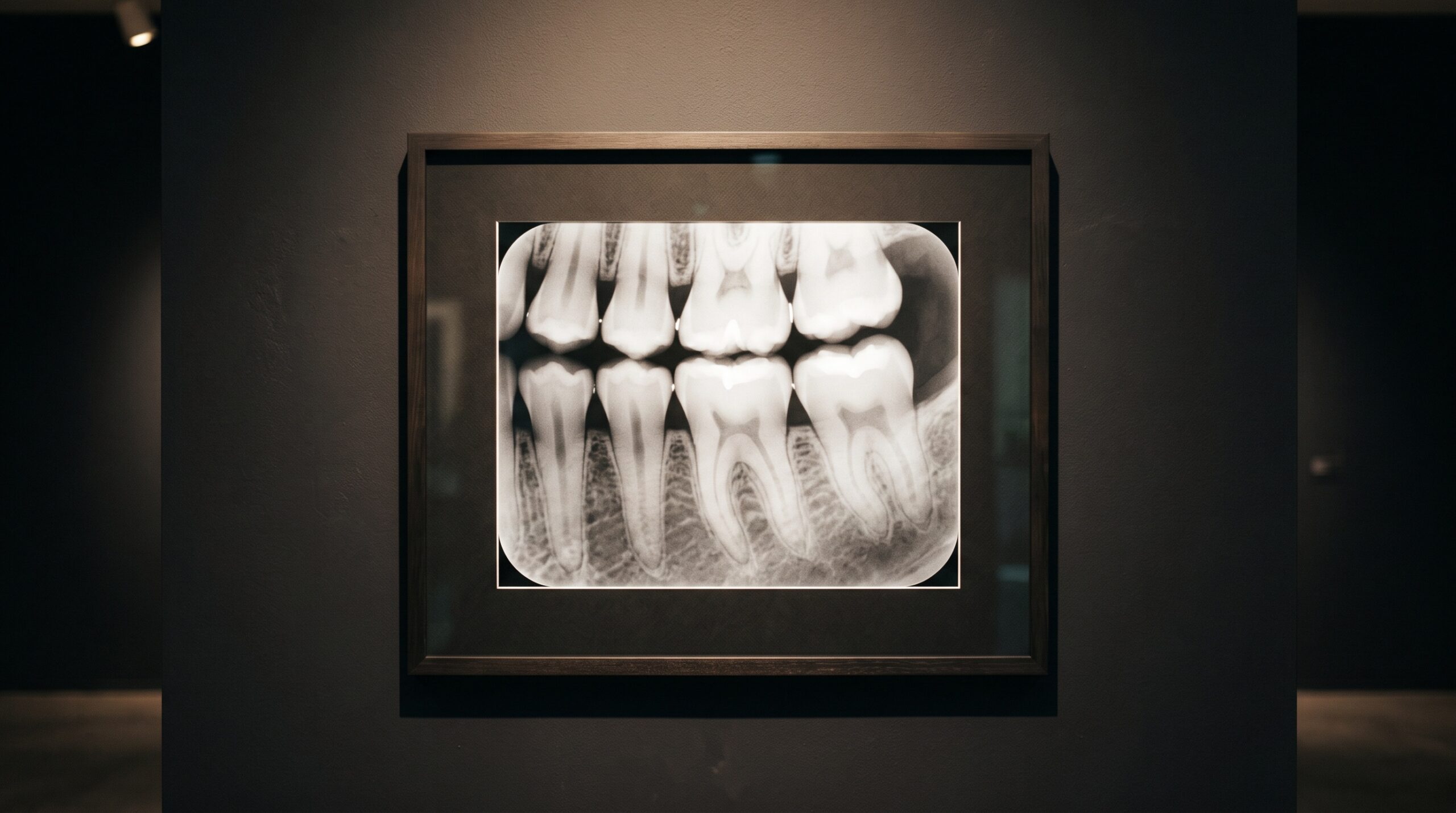
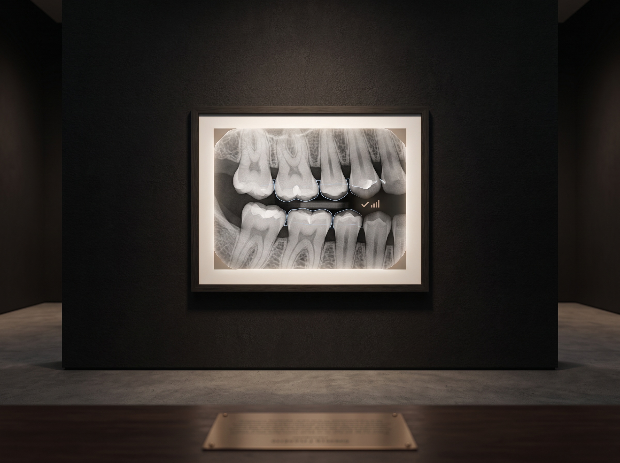
Of all the small exposures a practice makes in a day, the premolar bitewing is the one most often quietly wrong. It looks simple, a horizontal film of the back teeth caught in gentle occlusion, and yet it carries a demand the periapical never does: it must render the proximal surfaces of several teeth, from the distal of the canine through the first molar, as cleanly separated walls, each contact point opened into a thin bright line of space. When the contacts overlap, the single most important thing a bitewing exists to show, incipient decay hiding between the teeth, vanishes into a smear of superimposed enamel. The margin for getting it right is narrow, measured in a few millimetres of receptor position and a few degrees of beam, and the craft of the premolar bitewing is the craft of respecting that margin every time.
The One Job of a Bitewing
A bitewing is a purpose-built instrument, not a general view. Its reason for existing is the interproximal surface, the vertical wall where two adjacent teeth touch, because that is where caries begins unseen and where a clinical eye cannot reach. To serve that purpose the image must do one thing above all: open the contacts, projecting each pair of touching surfaces as two distinct edges with daylight between them rather than one overlapped shadow. Everything else about the technique, where the receptor sits, how the beam is aimed, how the patient bites, is in service of that single geometric goal. A bitewing that shows beautiful bone levels and perfect density but closes the premolar contacts has failed at the only task that distinguishes it from any other film. Understanding the view this way reframes placement as not a matter of comfort or habit but of geometry: the proximal walls of the premolars must line up with the beam, and the receptor must be where those walls will land.
Reading the Landmark: Where the Receptor Actually Belongs
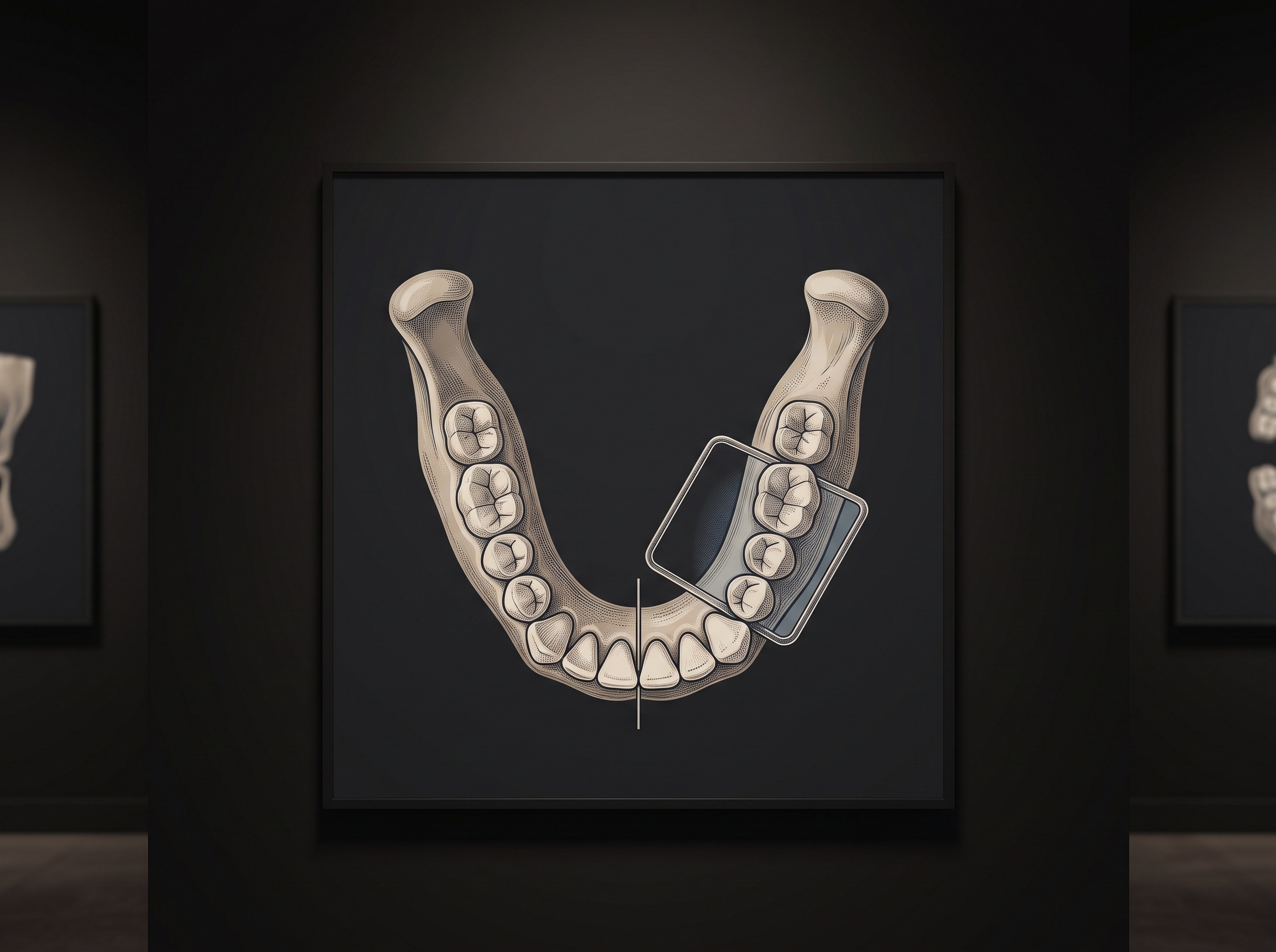
The premolar bitewing has a precise anterior landmark, and most overlapped films come from ignoring it. The front edge of the receptor should sit at the middle of the lower canine, far enough forward to capture the distal surface of that canine, because the contact between canine and first premolar is itself a site caries loves and a surface no periapical reliably opens. Set the tab too far back, a reflexive move to keep the patient comfortable, and that canine-premolar contact falls off the front of the film entirely; set it too far forward and the distal of the first molar, the other anchor the premolar view is meant to include, drops off the back. The correct placement threads both: front edge at the canine’s midline, which naturally carries the posterior edge to the distal of the first molar. On a tab film this means the tab is attached roughly over the first and second premolars and the patient closes gently with the receptor held against the lingual of the lower arch, the film angled slightly to follow the arch rather than fighting it. On a holder such as a Rinn-style bite-block system the landmark is the same; the device simply fixes the relationship between receptor and beam so the operator need only position the whole assembly to that canine-midline mark.
Why Premolars Overlap: The Curve Nobody Accounts For
Premolar contacts overlap for a reason rooted in anatomy, and naming it is the first step to defeating it. The dental arch curves, and the premolar region sits on the bend where the flat anterior segment turns toward the posterior. A beam aimed straight at the molars, or straight back along the patient’s sagittal line, strikes the premolar contacts obliquely, passing through the mesial surface of one tooth and the distal of its neighbour at an angle and superimposing them. The correction is horizontal angulation: the central ray must be directed through the embrasures, perpendicular to the proximal surfaces at that specific point on the curve, which for the premolars means aiming slightly more from the front than the molar region requires. This is why a single horizontal angle rarely opens an entire quadrant, and why the premolar and molar bitewings are separate exposures rather than one long film, a point worth remembering when weighing how many images a complete examination truly needs, as we discussed in reading the diagnostic question behind a full-mouth series against cone-beam CT. The beam must meet the curve where the curve actually is.
The Three Degrees That Separate a Film From a Retake
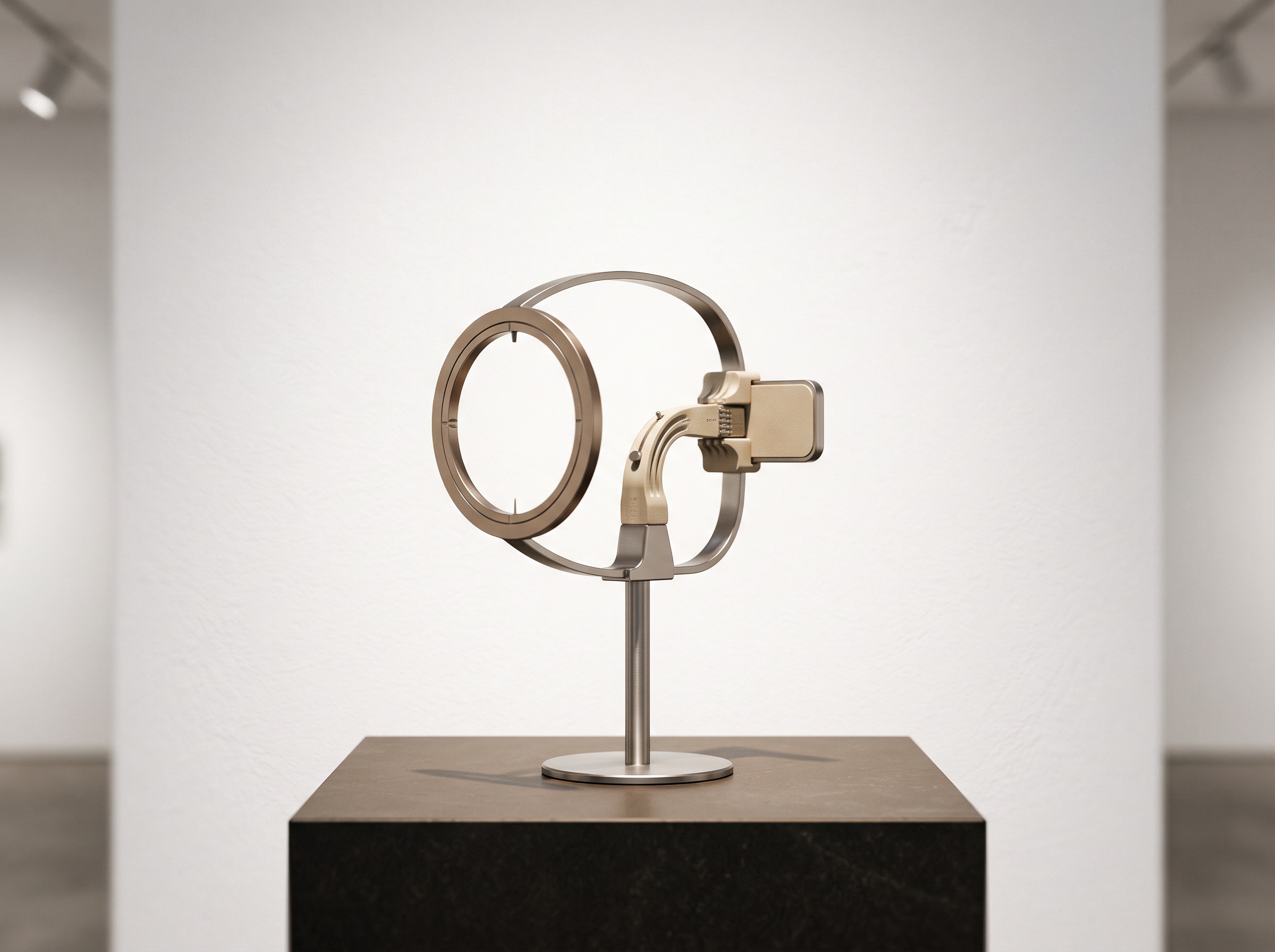
Horizontal angulation is unforgiving in the premolar region because the contacts are tight and the surfaces nearly parallel, so even a small error smears them. The discipline is to visualise the proximal planes before exposing, imagine the flat faces where premolar meets premolar and premolar meets molar, and align the beam to look straight down those planes rather than across them. With a positioning device that uses an external aiming ring, the ring does this work if the receptor is seated correctly; freehand, the operator must set the horizontal angle deliberately, resisting the instinct to simply point at the cheek. Vertical angulation matters less for contact opening but still governs whether the film records true bone levels or a distorted projection, and the two errors compound: a receptor placed well but shot at the wrong horizontal angle yields overlap, while a perfect angle on a poorly seated receptor clips the very contacts it was meant to show. Precision in both is what keeps the interproximal surfaces honest, the same attention to getting it right the first time that governs every low-dose discipline, including the careful technique we described for scaling dose and beam for the smallest patients.
The Retake Is a Dose, Not an Inconvenience
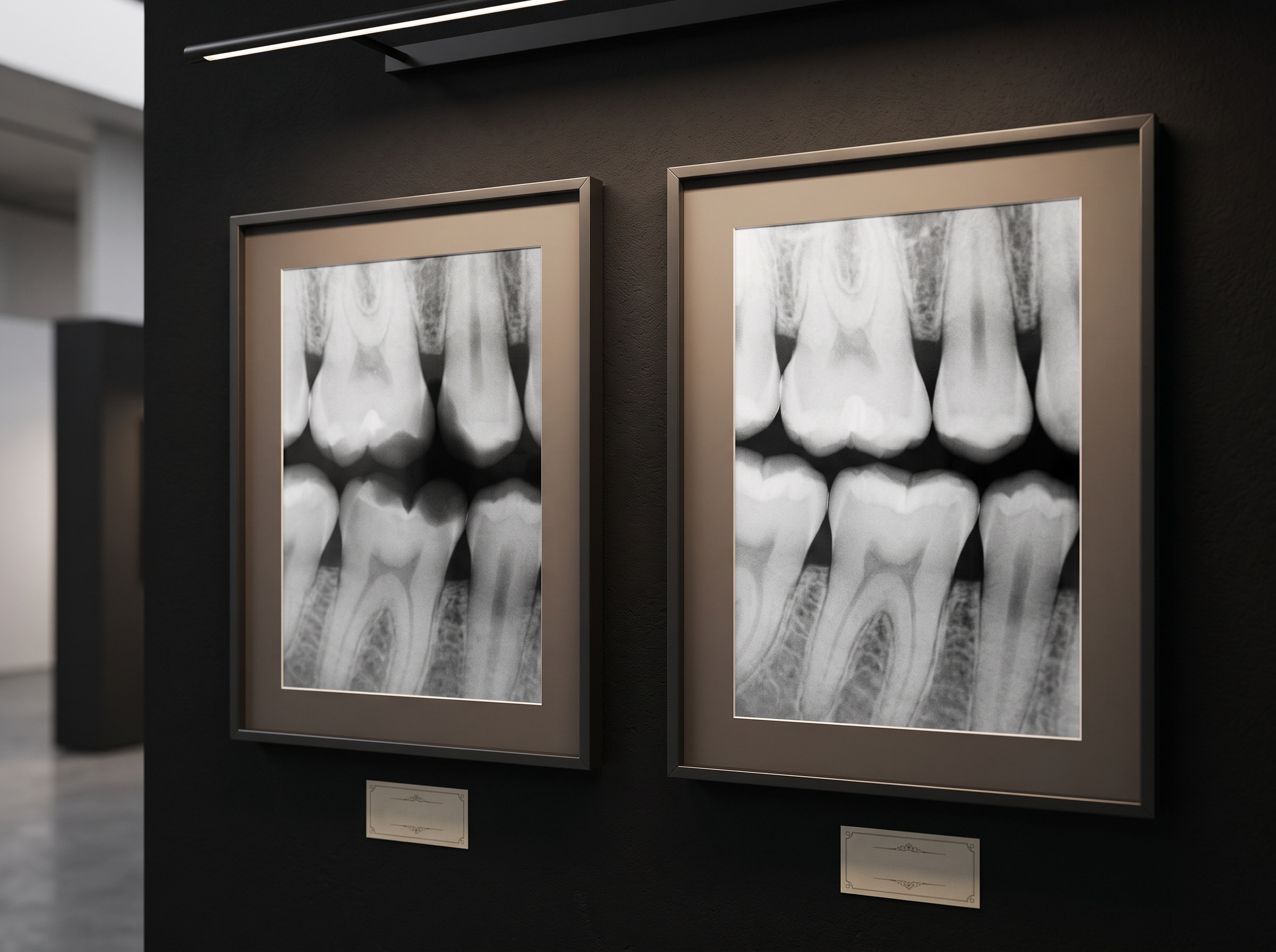
It is tempting to treat a closed-contact bitewing as a minor annoyance, a film to redo and move on from, but each retake is a second dose of ionising radiation delivered for an avoidable reason, and retakes concentrate in exactly the technique-sensitive views the premolar bitewing exemplifies. A practice that tracks its retakes, and few track them honestly, will usually find overlapped posterior contacts near the top of the list. The remedy is not more exposures but better first exposures: a reliable landmark, a deliberate horizontal angle, and a receptor holder that removes guesswork from the beam-to-film relationship. The same vigilance that catches a projected artifact before it is mistaken for pathology, the discipline we set out in recognising and preventing the phantom images that haunt a panoramic, applies to the quieter failure of the overlapped bitewing: the image that is not wrong so much as useless, and worth a retake only because the first attempt skipped a step. Reducing retakes is the most direct dose reduction a practice controls, because it is the one failure entirely within the operator’s hands.
Building the Habit Into the Hand
Technique that depends on inspiration fails under the pressure of a full schedule; technique built into habit survives it. The premolar bitewing rewards a fixed routine: seat the receptor with its front edge at the canine midline, confirm the distal of the canine and the distal of the first molar are both within the field, follow the arch rather than forcing the film flat against resistant anatomy, and set the horizontal beam to look straight through the premolar embrasures before pressing the button. A positioning holder turns much of this into mechanics, which is why consistent bitewings and consistent holders tend to arrive together. The operator who internalises the landmark stops thinking about it consciously and simply places the film correctly, the way a photographer stops calculating exposure and simply sees it, which is where craft in clinical imaging always ends up: a repeatable, unglamorous reliability that produces a diagnostic film on the first try, nearly every time.
Future Developments
The premolar bitewing will not disappear, but the tools around it are quietly tightening its tolerances. Positioning systems with integrated external aiming rings continue to shrink the space for horizontal error, and solid-state sensors with real-time preview let an operator see an overlapped contact before the patient has left the chair, collapsing the feedback loop that once meant a second visit. Artificial-intelligence quality checks now entering the operatory can flag a closed contact or a clipped landmark the instant an image is captured, turning the silent retake into an immediate correction and, over time, into a teaching signal that trains the hand. None of it replaces the fundamentals; a sensor that warns of overlap still relies on an operator who knows why premolars overlap and where the receptor belongs. The craft remains what it has always been, the patient discipline of meeting the curve of the arch with a well-aimed beam, so that the thin bright line between two teeth, the small space where early decay confesses itself, is there to be read.
No Comments