


November 2, 2026 The Smallest Patient: Scaling Dose, Beam, and Technique for Pediatric Dental Imaging
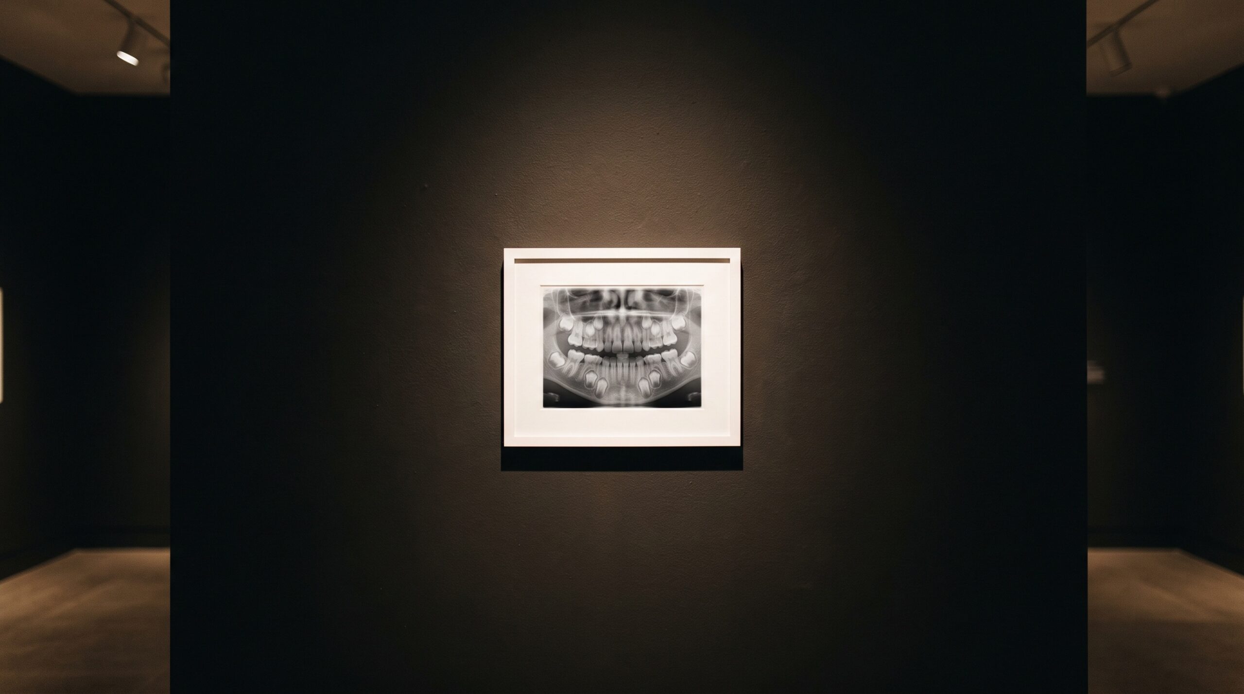
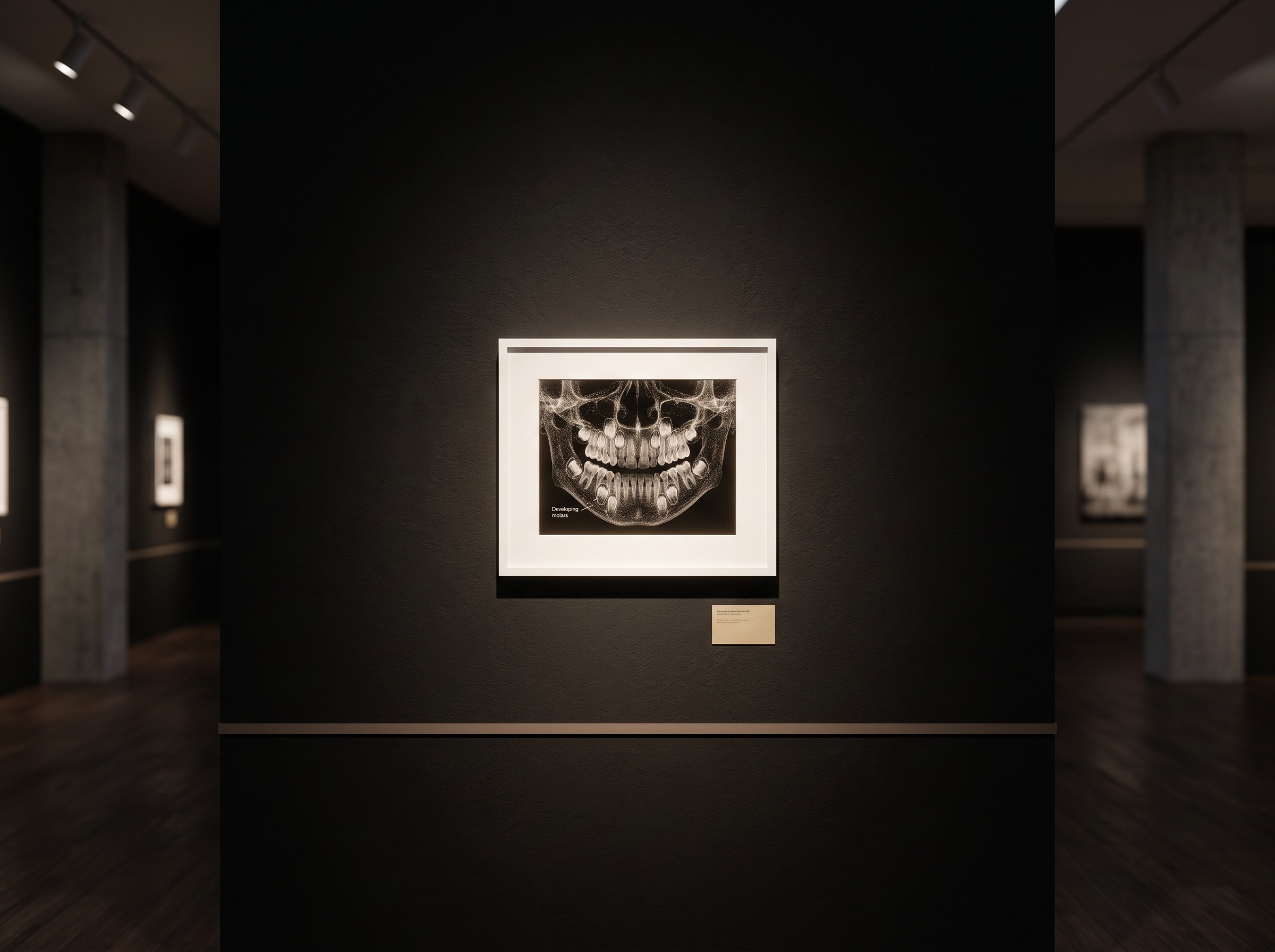
In a gallery of dental images, the pediatric radiograph is the smallest canvas and the most exacting to make. The anatomy is in motion, literally and developmentally: deciduous teeth loosening, permanent crowns rising beneath them, a jaw that will not look the same in a year. The temptation is to treat a child as a scaled-down adult and simply take the same pictures at a smaller setting. That instinct is wrong at almost every level. A child’s tissues are more sensitive to radiation, their bodies have more decades ahead in which a harm could surface, and their cooperation cannot be assumed. Imaging the youngest patients is not a lesser version of the craft. It is its most disciplined form, where every decision to expose must be earned and every exposure made as small as the diagnosis allows.
Why a Child Is Not a Small Adult
The physics of why children deserve special care is not sentimental; it is cellular. Rapidly dividing tissues are more radiosensitive, and a child’s growing bones, salivary glands, thyroid, and developing dentition are precisely the structures a dental beam passes near. Layered on top of that biological sensitivity is a simple matter of time: radiation-induced risk is stochastic and can take decades to manifest, and a seven-year-old has far more of those decades remaining than a seventy-year-old. The same absolute dose therefore carries a different lifetime weight depending on the age of the patient who receives it. None of this argues against imaging children when it is needed; untreated disease is its own harm. It argues for deliberateness, for treating each pediatric exposure as a considered act rather than a default.
The Discipline of Selection
The first and most powerful dose reduction is the image never taken. Routine radiographs on a fixed calendar, applied to every child regardless of risk, are the habit that modern guidance has worked hardest to retire. The shared position of the major bodies is that imaging should follow the individual child’s clinical findings, caries risk, and stage of development, not a schedule. A low-risk child with closed, caries-free contacts does not need bitewings as often as a high-risk child with proximal lesions, and no young patient needs a full survey simply because the chart has a blank field. This is the same stewardship we described in the ethic of the exposure and the discipline of imaging in moderation, sharpened by the fact that the patient is a child. And when an image is warranted, the next question is which one: the restraint of choosing the diagnostic question before the modality matters more, not less, when the subject is small.
Shaping the Beam, Shrinking the Dose
Once an exposure is justified, the craft turns to the equipment, and here a handful of unglamorous choices do most of the work. The pediatric-imaging safety movement, best known through the dental arm of the Image Gently campaign, distills decades of guidance into a short, practical discipline: select images for individual need, use the fastest available digital sensor or film speed, collimate the beam tightly to the area of interest, and protect the patient with a thyroid collar. Of these, rectangular collimation is the quiet giant. A conventional round beam irradiates far more tissue than a sensor actually captures; shaping that beam to the rectangle of the receptor can cut the exposed area several-fold, and with it the dose, at no cost to the diagnostic image. Child-size exposure settings matter too, because a smaller patient attenuates less and needs less, and a machine left on an adult preset over-exposes a child by default. None of these are new technologies. They are the difference between equipment that is merely present and equipment that is actually used well.

The Three-Dimensional Question
Cone-beam computed tomography has a place in pediatric dentistry, but it is a narrower place, and the justification bar sits higher. The volumetric scan that answers an impaction, a supernumerary tooth, a cleft, or a complex trauma can be genuinely decisive, sparing a child repeated two-dimensional attempts that would never resolve the question. But a CBCT also delivers more dose than a bitewing, and in a radiosensitive patient that trade must be explicit. When the scan is justified, the craft is to shrink it: the smallest field of view that captures the problem, a dedicated low-dose or pediatric protocol rather than an adult default, and reconstruction that wrings diagnostic clarity from fewer photons. That last point is where the technology is genuinely helping, as we explored in the deep-learning reconstruction that lets a scan be taken at a lower dose. The principle is constant: for a child, a three-dimensional image is a deliberate exception, never a routine.
The Patient Who Moves
There is a part of pediatric imaging that no collimator can solve, and it is the most human. A frightened or fidgeting child produces motion blur, retakes, and the quiet pressure to rush, and a retake is simply a second dose. The craft here is behavioral: an unhurried explanation in a child’s own vocabulary, a parent positioned where the child can see them, a sensor holder sized for a small mouth, and the patience to let a nervous patient settle rather than forcing a frame that will have to be made again. The best dose-reduction tool in the pediatric operatory is often not a setting at all but a calm minute spent before the exposure, because the image taken well the first time is the image a child never has to repeat.
Future Developments
The trajectory of pediatric dental imaging bends, encouragingly, toward less: lower dose, tighter beams, and software that reconstructs a usable picture from a fainter signal than was ever possible before. Artificial intelligence is beginning to flag which children genuinely warrant imaging and to rescue diagnostic detail from low-exposure captures, and the hardware is slowly standardizing on the collimation and child-aware presets that guidance has urged for years. But the deepest development is cultural rather than technical. It is the growing conviction that the smallest patient deserves the most care, that a radiograph of a child is a considered act with consequences measured in decades, and that restraint is not a limitation of the craft but its highest expression. At this gallery, the images we most admire are the ones made with exactly enough light and no more, and nowhere is that ideal more worth honoring than in the pictures we make of the young.
Sources & further reading:
- Image Gently Alliance – Dental (The Alliance for Radiation Safety in Pediatric Imaging)
- FDA – Dental Radiographic Examinations: Recommendations for Patient Selection and Limiting Radiation Exposure
No Comments