


September 19, 2026 The Instrument Enters the Laboratory: A Donated Cone-Beam Scanner and the Craft of Studying What We See
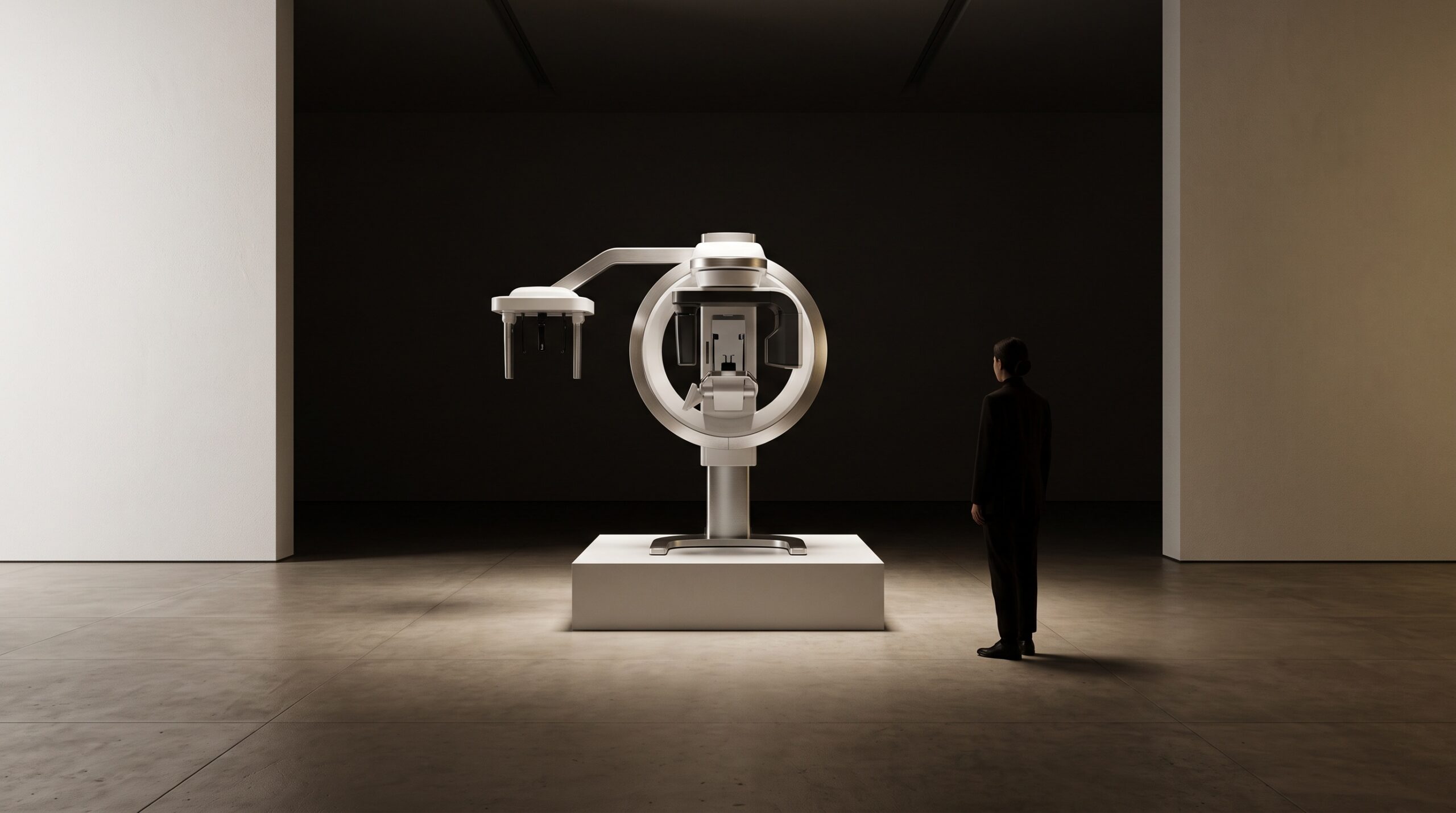
In late July 2026, Carestream Dental announced a strategic alliance with the ADA Forsyth Institute, and sealed it with a gift: a state-of-the-art cone-beam computed tomography system, donated to the institute’s clinical arm, ADA Forsyth Faculty Associates. It would be easy to file that under corporate philanthropy and move on. It is more interesting read as a quiet statement about where imaging is studied, and by whom – because when a manufacturer places a research-grade scanner inside one of the field’s leading laboratories, it is not simply giving away a machine. It is placing an instrument where the images it makes will be taken apart, questioned, and improved.
The Gift That Is Really an Instrument
The ADA Forsyth Institute is not a dental office. It is a leading independent, NIH-funded research organization devoted to the connections between oral health and the rest of the body, and its clinical facility exists to turn patient care and inquiry into the same act. Donating a cone-beam scanner to a place like that is less like furnishing an operatory and more like endowing a telescope. The equipment will certainly image patients – but it will also be the object of protocols, comparisons, and measurements that a busy private practice has neither the time nor the mandate to run. Carestream has done this before, placing systems at university dental colleges and teaching institutes over the past decade, and the logic is consistent: the fastest way to learn what an imaging system can really do is to hand it to the people whose job is to find out.
Why a Research Clinic Wants a Cone Beam
To understand the significance, it helps to remember what cone-beam computed tomography actually is. Where a conventional radiograph flattens the jaws into a single plane – overlapping roots, superimposed bone, and hidden canals collapsed into one shadow – a CBCT sweeps a cone of X-rays and a flat-panel detector around the head in a single rotation and reconstructs a true three-dimensional volume from the hundreds of projections it captures. That volume can be sliced in any plane, rotated, and measured, which is precisely why it reveals the anatomy a flat film cannot: the extra canal, the fused or dilacerated root, the exact relationship of a lesion to the nerve. We have written before about the hidden architecture of a tooth that cone-beam CT reveals but a flat radiograph conceals, and about the frontier beyond it – spectral, dual-energy CBCT that can tell bone from metal by reading two X-ray energies at once. A research clinic wants a scanner of this caliber because the questions it can ask – about dose, about artifact, about what the reconstruction is really measuring – can only be asked with equipment good enough to trust.
From the Bench to the Operatory
The most consequential thing about a research-grade scanner in a research clinic is the loop it closes. The acquisition protocols that lower dose without sacrificing diagnostic yield, the artifact-correction methods that clear the streak a metal restoration throws across a slice, the reconstruction algorithms that sharpen bone margins, and increasingly the annotated datasets that teach dental AI to read a volume honestly – these are not born in the operatory. They are validated on equipment held to a standard higher than daily practice requires, by people whose task is to be skeptical of their own images. Then they travel outward, as firmware, as guidelines, as the default settings on the machine a private practice buys three years later. A scanner in a laboratory is, in that sense, an investment in the images every clinician will eventually make without ever knowing where the refinement came from.
The Discipline Behind the Dose
There is a temptation to treat a powerful imaging system as a reason to image more. A serious research institute is exactly the place where the opposite discipline gets built – the evidence for when a three-dimensional scan is genuinely justified and when a simpler exposure, or none at all, is the better answer. Cone-beam CT delivers more radiation than a conventional radiograph, and the case for using it rests on the diagnostic value it adds in a specific situation, not on its availability. That is the ethic behind the discipline of restraint and the art of the radiograph not taken, and it is the kind of judgment that laboratories with the equipment and the mandate are positioned to formalize into selection criteria the rest of the profession can lean on. A donated scanner, used well, produces better guidance about imaging less – a paradox only in appearance.
Imaging as an Object of Study
PatientGallery has always held that a radiograph is not only evidence about a patient but an artifact worth regarding in its own right. A research clinic makes that literal. There, the reconstruction is examined not just for what it says about a mouth but for what it reveals about the imaging process itself – the resolution it truly resolves, the noise it introduces, the way its grayscale maps to real tissue density, the places where the algorithm has guessed. It is the difference between viewing a painting and studying it: the conservator who reads the under-drawing, the pentimenti, the ground beneath the paint, learns things the ordinary visitor never sees. A scanner in a laboratory turns every volume into that second kind of viewing, and the field is better for having somewhere the image is interrogated as closely as the anatomy.
Future Developments
What an alliance like this accelerates is rarely a single dramatic breakthrough; it is the slow, compounding refinement that eventually feels like it was always there. Expect the work to push on the frontiers that matter most – lower-dose acquisition that widens the range of cases where a 3D view is defensible, photon-counting and spectral detectors that read tissue composition rather than mere density, and standardized, well-annotated datasets that let dental AI be trained on images a research institute is willing to vouch for. Each of those advances will arrive quietly in ordinary operatories, embedded in the next generation of machines. The gift of a scanner to a laboratory is, in the end, a bet on that trajectory: that the surest way to improve the image on every clinician’s screen is to put a very good instrument in the hands of the people whose calling is to understand exactly what it sees. At PatientGallery, that has always been the point – that imaging advances not only as technology but as knowledge, and that the two are, properly understood, the same craft.
Sources & further reading:
- Carestream Dental, ADA Forsyth Institute announce strategic initiative
- ADA Forsyth Institute | American Dental Association
No Comments