



August 18, 2026 The Placard Beside the Picture: How Structured Reporting Makes a Dental Radiograph Legible
Walk through any gallery and you learn quickly that the picture is only half of what you receive. The other half is the small placard on the wall beside it – the title, the medium, the line or two that tells you where to look and what you are seeing. A masterwork with a careless label is diminished; a modest study with a precise one is elevated. The radiograph lives under the same law. The image captures the tooth in exquisite grayscale, but the diagnosis is not complete until someone writes down what the image says, and the form that writing takes turns out to matter far more than the profession has generally admitted. A new comparative study puts a number on it, and the number is not subtle.
The Report Is the Other Half of the Image
A periapical radiograph is one of dentistry’s most concentrated instruments: a high-resolution portrait of a single tooth and the bone around it, capable of revealing root morphology, periapical lesions, bone levels, endodontic status, and restorations that no clinical examination can show. We have written at length about how much craft goes into making that image faithful – the true measure of what a sensor can resolve, and the calibrated screen on which it is finally read. But once the image is seen, its findings must be set down in words, and those words are what travel: to the next clinician, into the record, before a board, across a referral. The report is the durable artifact. The image may never be looked at again; the report is read every time.
Narrative Prose and Its Quiet Failures
For as long as there have been radiographs, dentists have described them the way one describes anything – in free-flowing prose. Narrative reporting is flexible and familiar; it lets a clinician say exactly what they notice in whatever order it occurs to them. That freedom is also its weakness. Free text is uneven from one practitioner to the next and, worse, from one busy afternoon to the next in the same practitioner. It omits. A finding not written is not merely unrecorded but effectively unseen by everyone downstream, and narrative prose gives an omission nowhere to show itself – a report that never mentions the furcation reads exactly like one where the furcation was checked and found sound. Across different clinicians and institutions, standardization all but vanishes, and with it the reproducibility that quality assurance, medico-legal clarity, and interdisciplinary communication all quietly depend upon.

What a Structure Actually Does
Structured reporting proposes a different discipline. Rather than a blank page, the reader works through a predefined template: named categories, standardized terminology, sometimes decision trees that carry the eye deliberately from root apex to crestal bone to restoration margin. The gain is not eloquence – structured reports rarely read like fine writing – but coverage and consistency. Every field is a question that must be answered, so the checklist itself becomes a guard against the thing free prose does worst: silent omission. The empty field is the whole point. Where a narrative simply moves on, a template leaves a labelled blank that announces a finding was not addressed. It is the same principle that lets a trained eye catch an illusion that mimics decay at the tooth’s neck – a fixed method of looking is what keeps attention from skating past the places it prefers to skip.
The Study That Measured It
Until recently this was an argument from principle. A 2026 comparative evaluation from the radiology department of the University Hospital at LMU Munich, published in Frontiers in Dental Medicine, turned it into an argument from evidence. The researchers took fifty narrative reports of intraoral periapical radiographs and, for each one, built a corresponding structured report from a standardized dental template. Two independent, experienced dentists then judged both versions of every case against nine parameters – completeness, information extraction, level of detail, logical sequence, trustworthiness, linguistic quality, clarity, therapeutic decision-making, and overall assessment – without the advantage of knowing which format was meant to win.
The structured reports were rated significantly higher – at a stringent level of statistical significance – on completeness, information extraction, detail, trustworthiness, linguistic quality, clarity, and overall evaluation. In other words, on nearly every axis that governs how well a reader can extract and trust what the image holds, the fixed structure beat the free narrative, and it did so decisively rather than marginally.
The Honest Exception
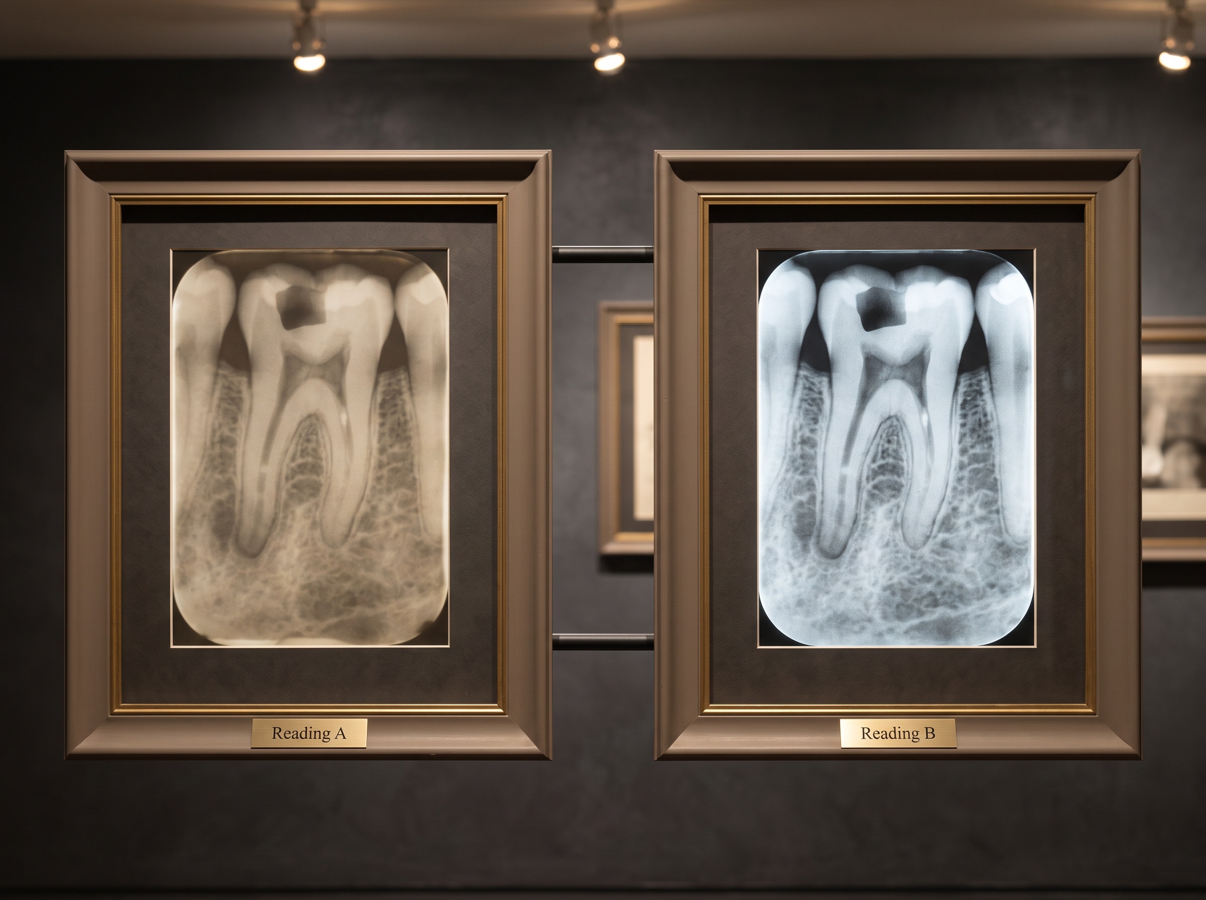
One result deserves its own paragraph, because it keeps the finding honest. On two parameters – therapeutic decision-making and whether the report carried enough information to plan treatment – the study found no significant difference between the two formats. Both the narrative and the structured report generally let the clinician arrive at the right call. This is not a weakness in the case for structure; it is the measure of its real value. Structured reporting did not make the two experienced readers better diagnosticians on these cases – they were already good enough to reach the correct decision from either format. What it improved was the legibility of that decision to everyone who was not in the room: the completeness of the record, the clarity of the communication, the trust a second reader can place in the first. The image was already saying the right thing; structure made sure the words around it said it clearly, and said all of it. That distinction – a faithful signal versus a faithfully communicated one – is the same one that separates a good radiograph from an over-processed image that flatters the eye while quietly deceiving it.
A Growing Consensus
The periapical study did not arrive alone. A companion investigation in the same journal examined structured reporting of dental panoramic images in a hospital radiology setting and reached a strikingly parallel conclusion: structure significantly enhanced report quality, clinical decision-making, and communication. Two different intraoral and extraoral modalities, two independent studies, one direction of travel. In medical radiology the case was settled some time ago – structured reporting has long been shown to improve the clarity, completeness, and reproducibility of documentation – and dental radiology, where its adoption has lagged, appears to be arriving at the same place. The evidence is beginning to converge on a conclusion that is almost aesthetic in nature: a disciplined frame serves the work better than an unconstrained one.
Future Developments
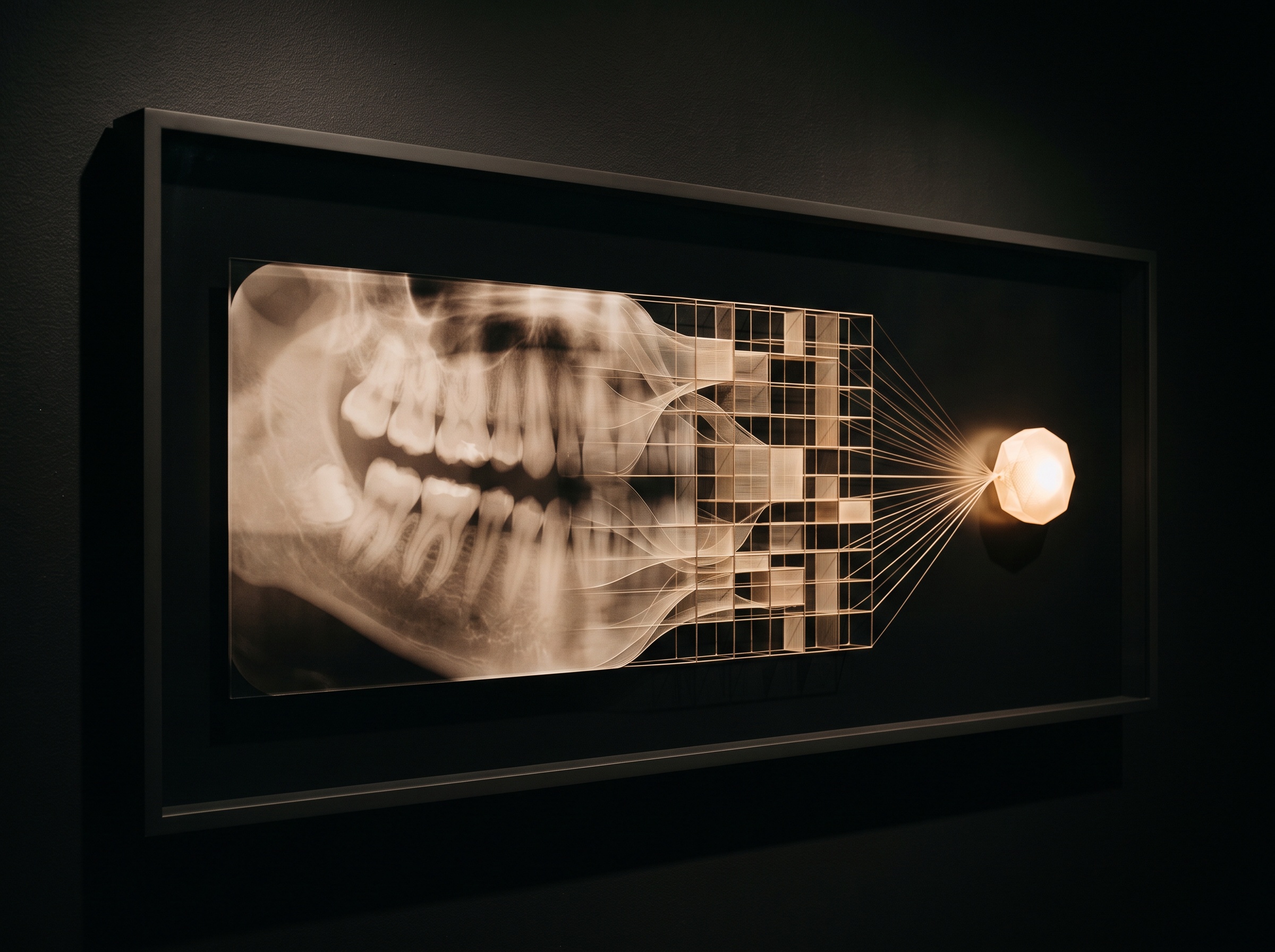
The most consequential argument for structure is not about the human reader at all. A narrative paragraph is opaque to a machine; a structured report, with its named fields and standardized terms, is already halfway to being data. As clinical decision-support systems and diagnostic AI move into the operatory, the report becomes the interface between a human’s reading of an image and a computer’s – and only a structured one can be parsed, audited, aggregated across a practice, or fed forward to a model without a translator in between. The near future likely holds templates that populate themselves as an algorithm pre-reads the radiograph and the clinician confirms or corrects each field, marrying the machine’s tirelessness to the human’s judgment. What the LMU study quietly demonstrates is that the groundwork for that future is not exotic technology but something a practice can adopt tomorrow: the decision to describe an image in a fixed, complete, consistent form. The picture has always deserved a good placard. It turns out the placard can be measured, and the measurement says to make it a careful one.
Sources & further reading:
Sorry, the comment form is closed at this time.