


November 21, 2026 CBCT at Panoramic Dose: How Pulsed Exposure and Beam Filtration Are Closing the 3D Radiation Gap
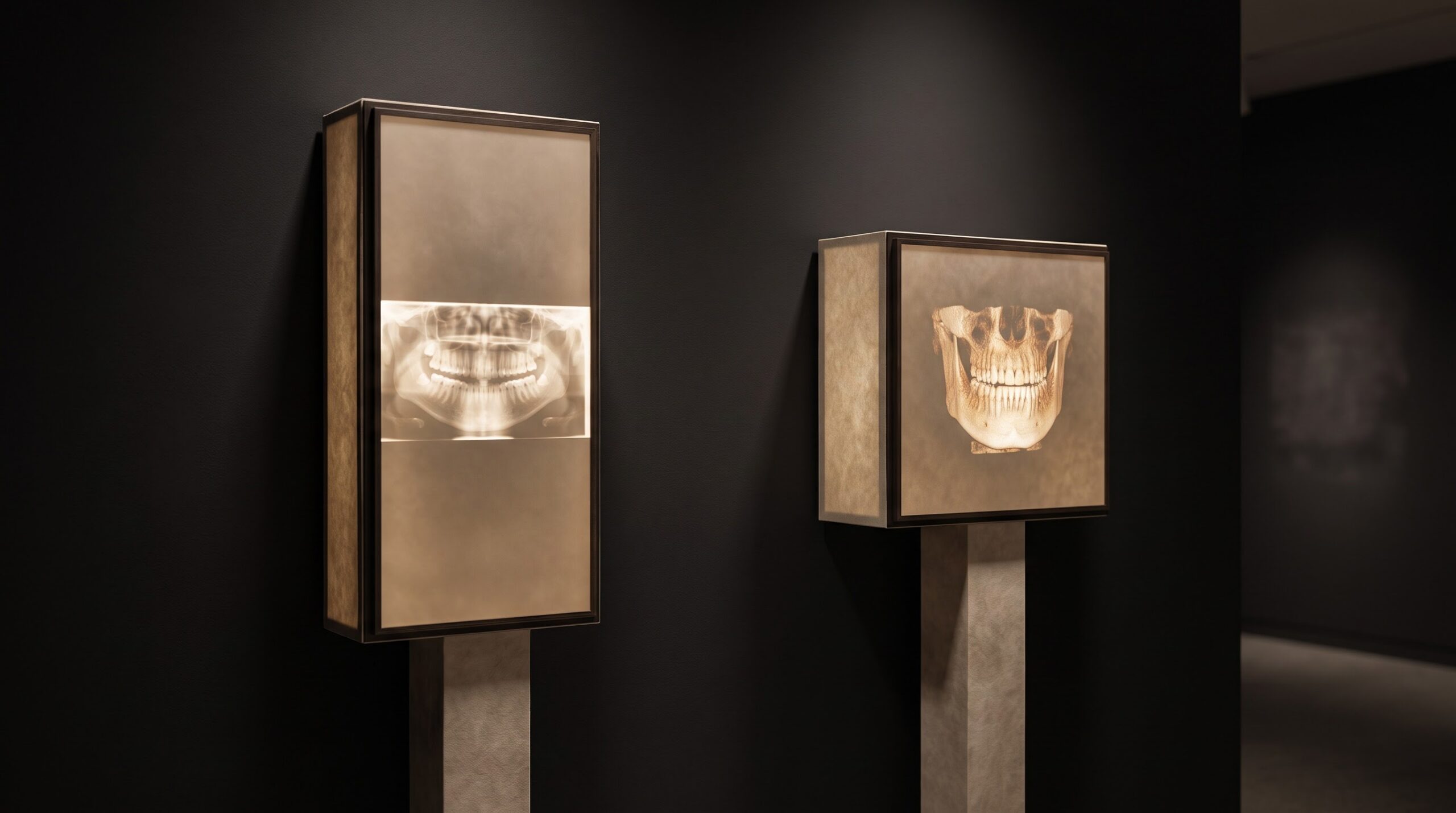
For most of the time that cone-beam computed tomography has lived in the dental operatory, it has carried a quiet asterisk. The three-dimensional view it offers is extraordinary — root canals traced in their true curvature, impacted teeth placed exactly against the nerve, bone volume measured before an implant — but the price has always been radiation. A panoramic radiograph sweeps the whole jaw for something on the order of ten to thirty microsieverts; a generous field-of-view CBCT could ask for many times that. The clinician’s decision was therefore framed as a hard trade between information and exposure. What the 2026 generation of hardware is doing, undramatically and from several directions at once, is dissolving that trade. The gap between a cone-beam scan and a panoramic exposure is narrowing to the point where, for a small targeted study, the two can begin to share the same order of dose.
None of this comes from a single breakthrough. It is the compounding of four separate engineering decisions, each attacking the dose from a different angle, each modest on its own and formidable in combination. Understanding them is worth the effort, because the same levers the manufacturer builds into the machine are the ones the operator chooses to pull at the console.
Pulsing the beam: irradiating only when the image is captured
The first and largest lever is temporal. A cone-beam unit builds its volume from a few hundred flat projection images taken as the tube and detector rotate around the head. In an older continuous-exposure design, the X-ray beam stays on for the entire rotation — radiating steadily even in the gaps between the instants a frame is actually recorded. A pulsed acquisition switches the beam on only during those brief capture windows and leaves it dark between them. The number of projections, and therefore the information available to reconstruct the volume, is unchanged; what falls away is all the exposure delivered while no image was being formed.
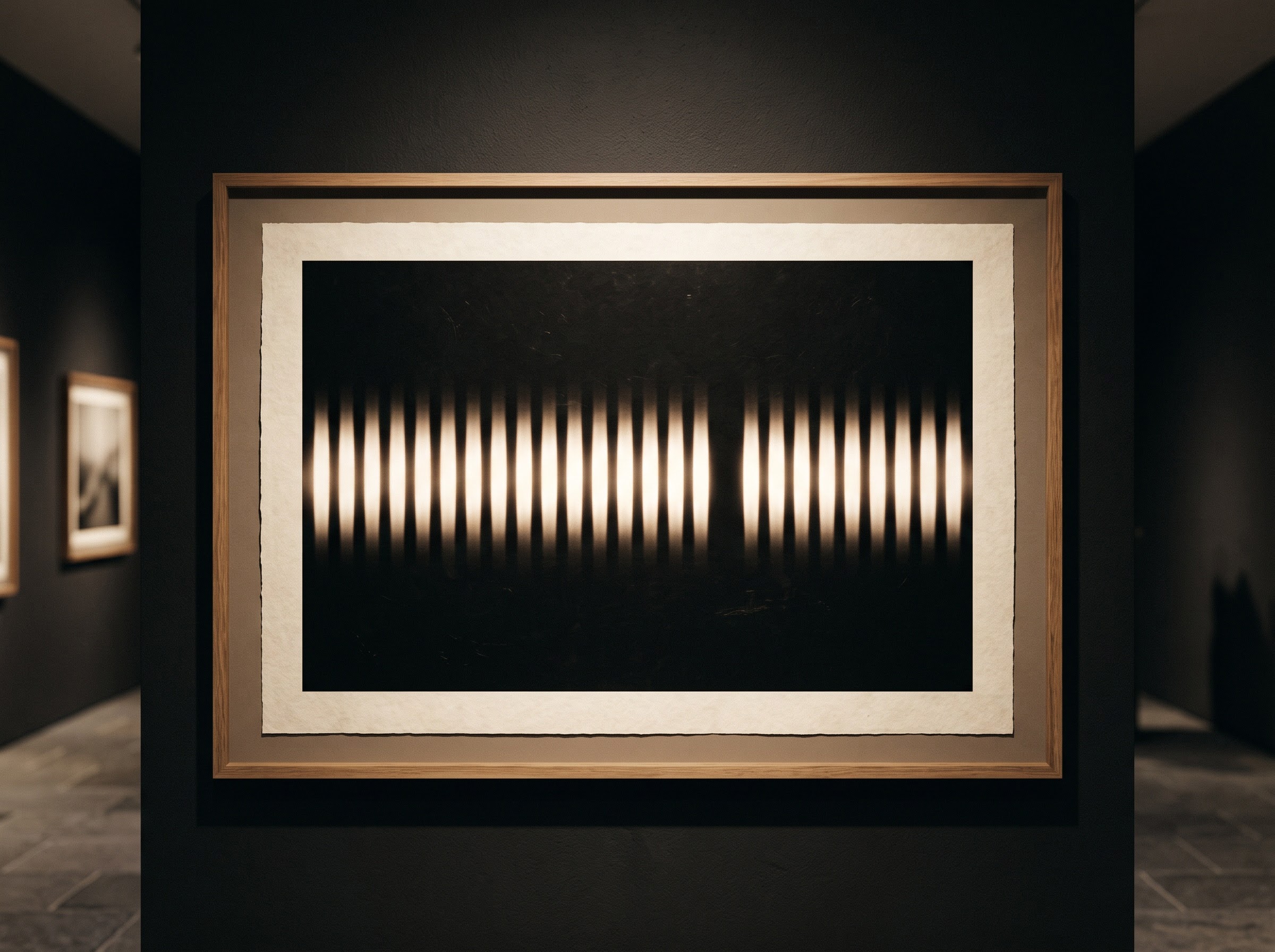
The effect is substantial. Depending on the frame rate and pulse width, the tube may be genuinely emitting for only a fraction of the nominal rotation time, and dose scales with that emitting time. Pulsing is the reason a scan that once felt expensive in radiation terms can now be acquired for a good deal less, with no visible cost to the geometry of the reconstruction. It is the cleanest kind of dose reduction — less exposure for the same diagnostic data.
Filtration: discarding the photons that only cost dose
The second lever shapes the beam itself. A raw X-ray spectrum leaving the tube is a mixture of energies, and its softest, lowest-energy photons are worse than useless for imaging: they lack the energy to pass through the jaw and reach the detector, so they contribute nothing to the picture while depositing their entire dose in the patient’s tissue on the way in. Added filtration — typically thin plates of aluminium and copper placed in the beam path — preferentially absorbs that soft tail before it ever reaches the face, hardening the beam into a cleaner, more penetrating spectrum.

The trade is favourable because what is removed was never doing diagnostic work. Heavier filtration raises the beam’s half-value layer and trims the skin dose markedly, at the cost of a tube that must work a little harder to maintain flux. For the patient this is close to a free lunch in dose terms, and it pairs naturally with the dose accounting that sites increasingly formalise through diagnostic reference levels and dose benchmarking, where every microsievert a protocol spends is measured against what comparable practices achieve.
Collimation: imaging the question, not the whole head
The third lever is spatial, and it is as much a clinical decision as an engineering one. CBCT dose is tied directly to the volume of tissue placed in the beam, so the single most reliable way to lower it is to irradiate less anatomy. Field-of-view collimation narrows the cone to cover only the region the clinical question concerns — a quadrant, a few teeth, a single surgical site — leaving everything outside that box in protective shadow. A small, tightly collimated field can carry a fraction of the dose of a full craniofacial scan while answering the question just as completely.
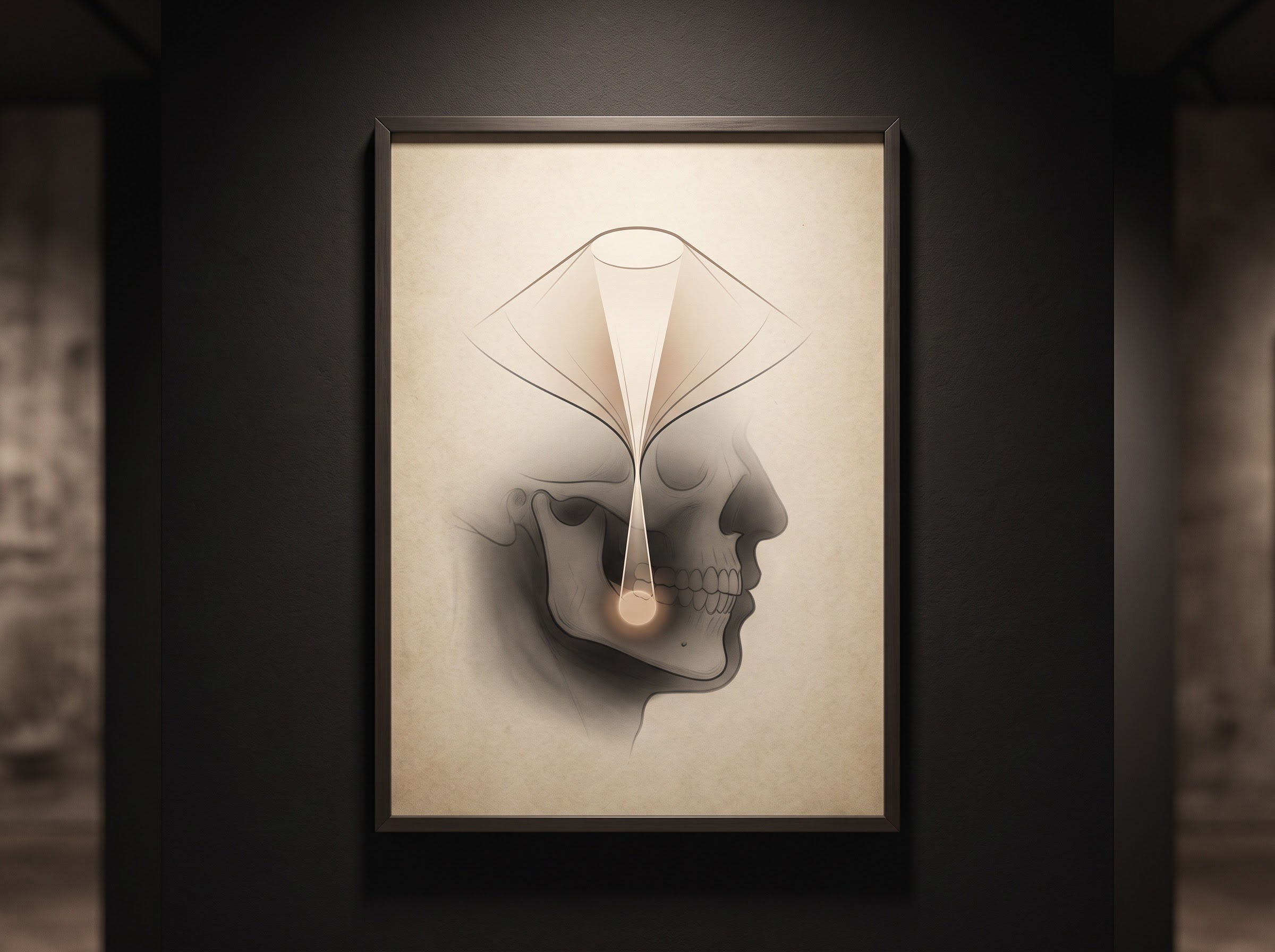
This is where dose reduction becomes a matter of discipline rather than hardware. Choosing the smallest field that still contains the diagnostic target is the operator’s analogue of the engineer’s filter plate, and it is inseparable from sound case selection — the same reasoning that governs when a full-mouth series serves better than a cone-beam volume at all. The machine offers the small field; the clinician has to be willing to use it, resisting the reflex to acquire more anatomy than the question requires simply because the detector can hold it.
Reconstruction: recovering the image the low dose gave up
Each of the first three levers lowers the number of photons reaching the detector, and fewer photons mean more noise — the grainy, mottled texture that can swallow a fine bone margin or a hairline root fracture. For years this noise penalty set a floor under how low dose could go before the images became diagnostically unreliable. The fourth lever raises that floor. Iterative reconstruction, and more recently deep-learning reconstruction, replace the fast but noise-blind mathematics of older algorithms with methods that model the physics of the acquisition and actively suppress noise while preserving edges.
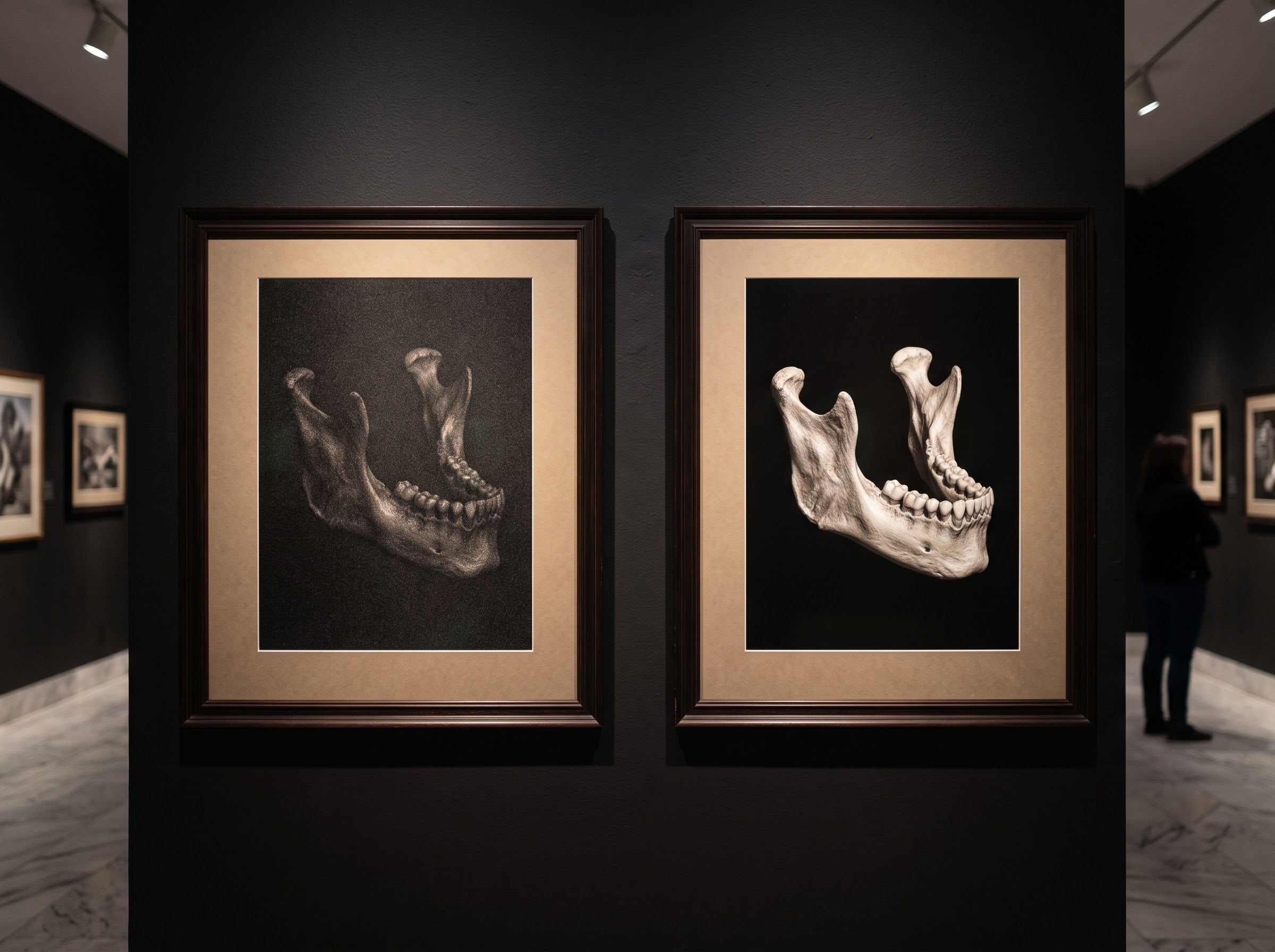
The practical consequence is that an acquisition deliberately starved of dose can be computationally restored to the clarity a diagnosis needs, decoupling image quality from raw exposure in a way that was impossible a hardware generation ago. The progress here has been rapid, and it is explored in its own right in our look at deep-learning reconstruction for low-dose dental CBCT. Reconstruction is the quiet partner of every other dose-saving measure: it is what makes aggressive pulsing, heavy filtration and tight collimation diagnostically safe rather than merely cheaper.
What the shrinking gap means at the chairside
Put together, these four levers change the shape of the clinical decision. The old framing — information versus dose, take the panoramic to be safe — assumed a fixed and punishing cost for the third dimension. When a small-field, pulsed, filtered, iteratively reconstructed scan approaches panoramic territory in dose, that assumption loosens. It does not vanish: a true one-to-one equivalence depends on the field size, the anatomy and the task, and a large craniofacial volume will always ask more than a quadrant study. The point is not that CBCT is now free of radiation concern but that dose has become a dial the clinician turns deliberately rather than a fixed toll paid for looking in three dimensions.
That shift places the emphasis exactly where it belongs: on justification and optimisation. The discipline is still to ask whether the three-dimensional view will change the treatment, and to accept the lowest dose that answers the question — the ALARA habit that governs every exposure, and that matters most acutely in the dose-sensitive protocols described in our piece on pediatric dental imaging and the craft of dose safety. The hardware has given the clinician room to say yes to the richer image more often; the responsibility is to keep saying it for the right reasons.
Future Developments
The direction of travel is clear, and it points at a quieter cone-beam still. Photon-counting detectors, already migrating from medical CT toward the maxillofacial scale, promise to wring diagnostic signal from each individual photon with an efficiency that today’s energy-integrating detectors cannot match, pushing the dose floor lower again. Reconstruction models trained on ever-larger image libraries will keep recovering clarity from thinner and thinner exposures, and task-adaptive protocols — where the machine proposes the field, frame rate and filtration best suited to the stated clinical question — will take more of the optimisation burden off the operator. The destination is a scan whose dose is shaped precisely to its purpose, no larger than the question demands. In the gallery of the dental image, the three-dimensional view is losing the shadow of radiation that once hung over it, and becoming simply another way of seeing — chosen for what it reveals, no longer feared for what it costs.
Sources & further reading:
No Comments