


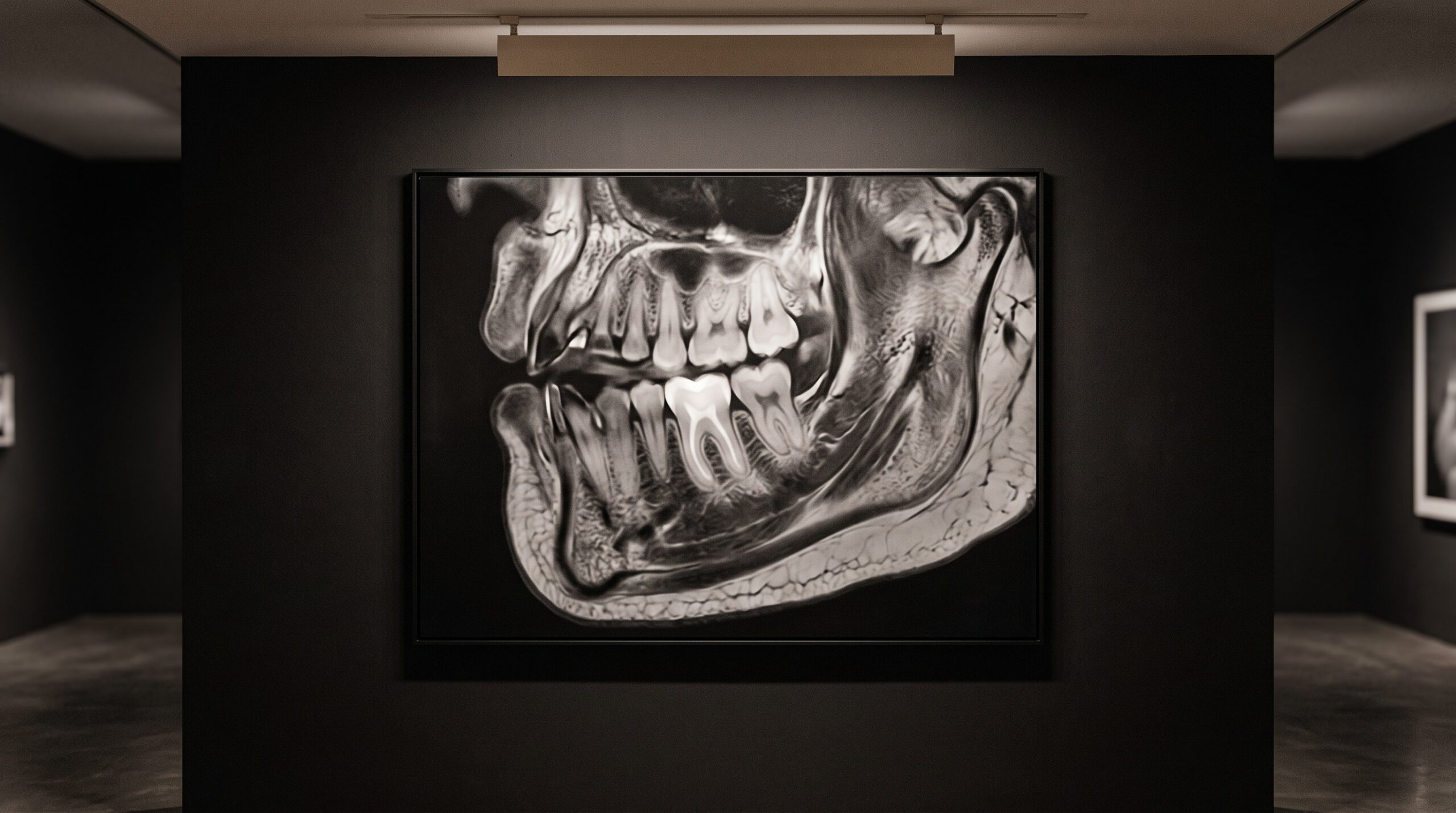
November 5, 2026 The Soft-Tissue Subject Arrives: A Dental-Dedicated MRI Clears the FDA, and the Gallery Gains a New Kind of Image
For as long as this gallery has hung radiographs on its walls, one subject has refused to sit for its portrait. The bone agreed to be photographed. The enamel, the restoration, the root and its canals, all of them rendered themselves obligingly in the grays of absorbed radiation. But the pulp inside the tooth, the inflamed ligament, the displaced disc of the jaw joint, the nerve threading its canal, these the x-ray could only imply, drawing their outlines by what surrounded them and leaving the thing itself unpictured. In March 2026 that absence acquired a remedy. A dental-dedicated magnetic resonance scanner, the MAGNETOM Free.Max Dental Edition, cleared the United States FDA on the strength of a completed clinical trial, and the soft-tissue subject that every radiograph has quietly omitted finally has an instrument built to render it.
From the Future Tense to a Cleared Instrument
The dental MRI has long been discussed the way one discusses any elegant possibility, in the conditional. We have written before about its promise, about a radiation-free way of seeing that seemed always a few years off. What changed in 2026 is grammatical as much as technical: the device moved from could to has. Developed by Dentsply Sirona together with Siemens Healthineers, the system did not arrive on a brochure but through the unglamorous discipline of a clinical trial, validated across multiple dental specialties and then cleared by a regulator whose entire purpose is to doubt. That provenance matters to a gallery that cares how an image is made. An unproven modality is a rumor; a cleared, trial-backed one is an instrument, and an instrument can be learned, critiqued, and eventually mastered as its own craft.
A Different Physics of Seeing
What makes the picture radically new is that it is not made of radiation at all. A radiograph, and its three-dimensional cousin the cone-beam scan, is a map of how densely tissue absorbs an x-ray beam; it is superb at hard structure precisely because calcified tissue stops photons. Magnetic resonance reverses the entire premise. It coaxes the hydrogen protons in water and fat to align in a strong magnetic field, nudges them with radio pulses, and listens to the faint signal they give back as they relax. Tissues rich in water and movement sing loudly; dense calcified bone, poor in free protons, stays comparatively dark. The result is an image whose strengths are the exact inverse of the radiograph’s. Where the x-ray excels at the mineralized and goes mute on the soft, MRI renders the soft tissue in rich tonal gradation and treats the hard shell almost as context. For the first time the two families of dental image are genuinely complementary rather than competing, each picturing what the other cannot.
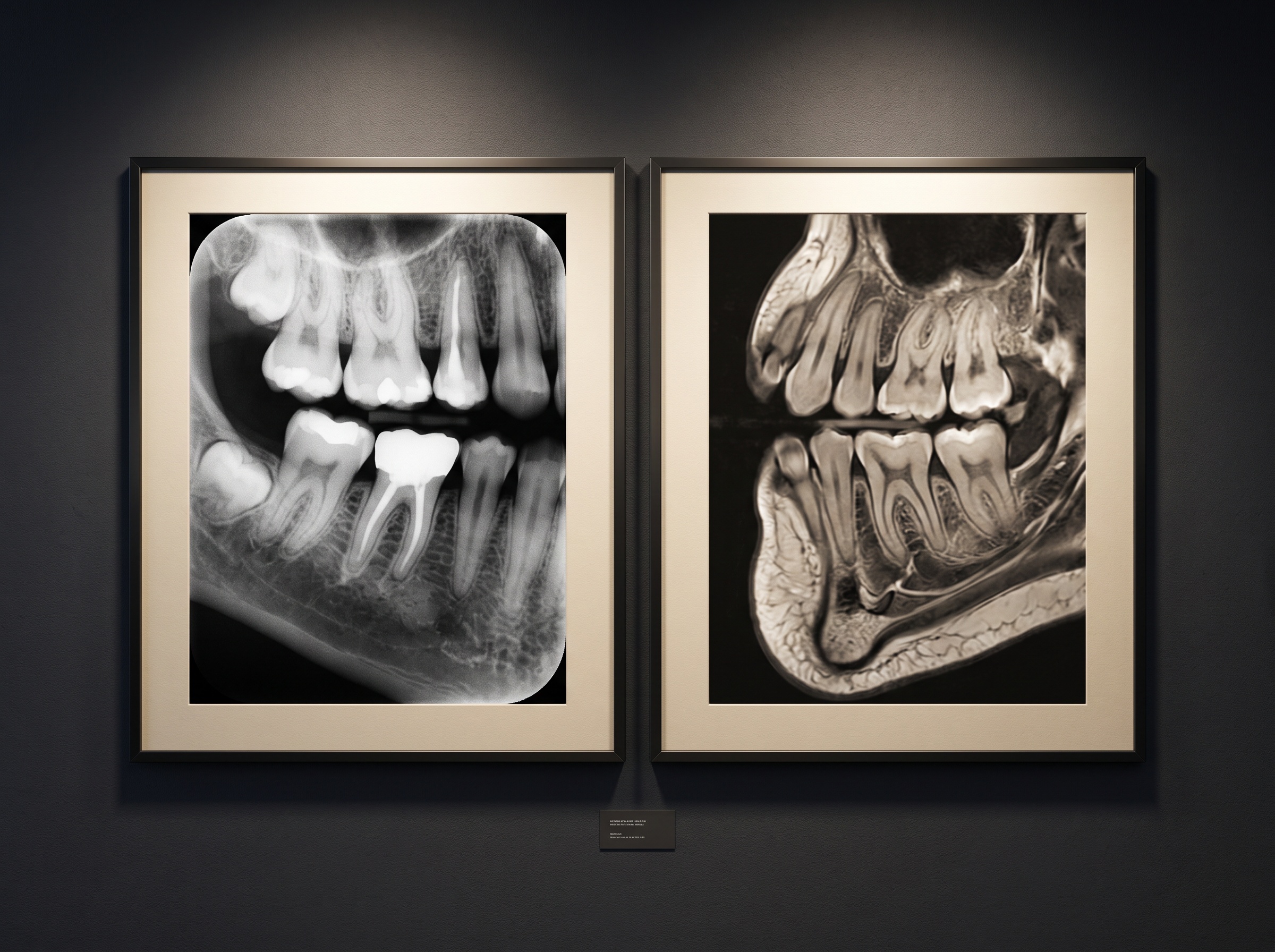
The Subjects That Were Always Missing
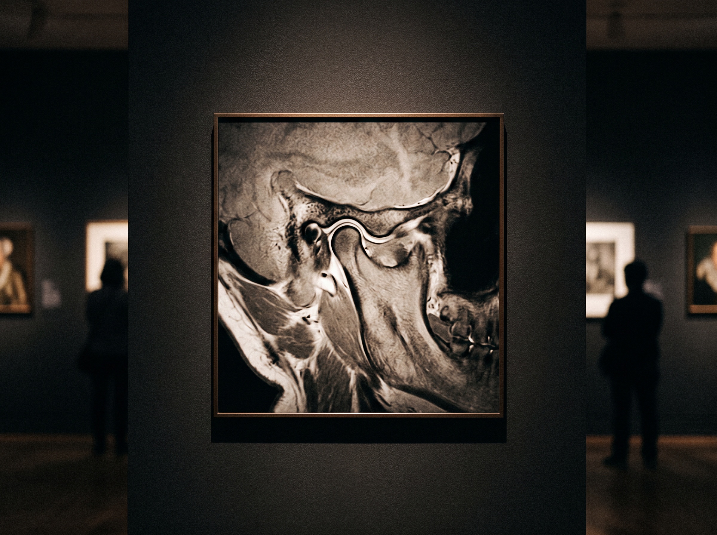
Consider what this opens to view. The dental pulp, long inferred only from the shape of the space it occupies, becomes a tissue one can actually assess. Inflammation in the periodontal ligament, the early biology of a lesion before it has carved itself into bone, the soft architecture of the nerve, all of these step out of implication and into the picture. Nowhere is the gain more obvious than at the temporomandibular joint, where the articular disc, a sliver of fibrocartilage invisible to any x-ray, is the very structure clinicians most need to see. That is terrain we have mapped before in discussing how MRI and CBCT together picture the joint that moves; what has changed is that the MRI half of that pairing is now a purpose-built dental device rather than a borrowed hospital scanner. The soft tissue of the airway, too, which a cone-beam scan can only outline as a column of air, gains a modality suited to the walls that define it, extending the kind of reading we described in the airway hidden inside the scan.
No Dose, and What That Changes
The quiet revolution underneath all of this is the absence of ionizing radiation. Every radiographic image a dentist makes is a negotiation with dose, a weighing of diagnostic yield against a small, real exposure, the discipline of asking whether a given picture earns its photons. An MRI image asks for none of that. There is no cumulative burden, no special caution for the young patient or the frequently imaged one, no justification to log against a sievert budget. This does not make MRI a replacement for the radiograph, which remains faster, cheaper, and unmatched for hard tissue and for the quick confirmation that fills a clinical day. But it reframes the whole question of when to look. When the thing you need to see is soft, the answer need no longer carry a radiation cost at all, which is a genuinely new freedom in a field that has always paid for its vision in dose.
Where the Modality Actually Fits
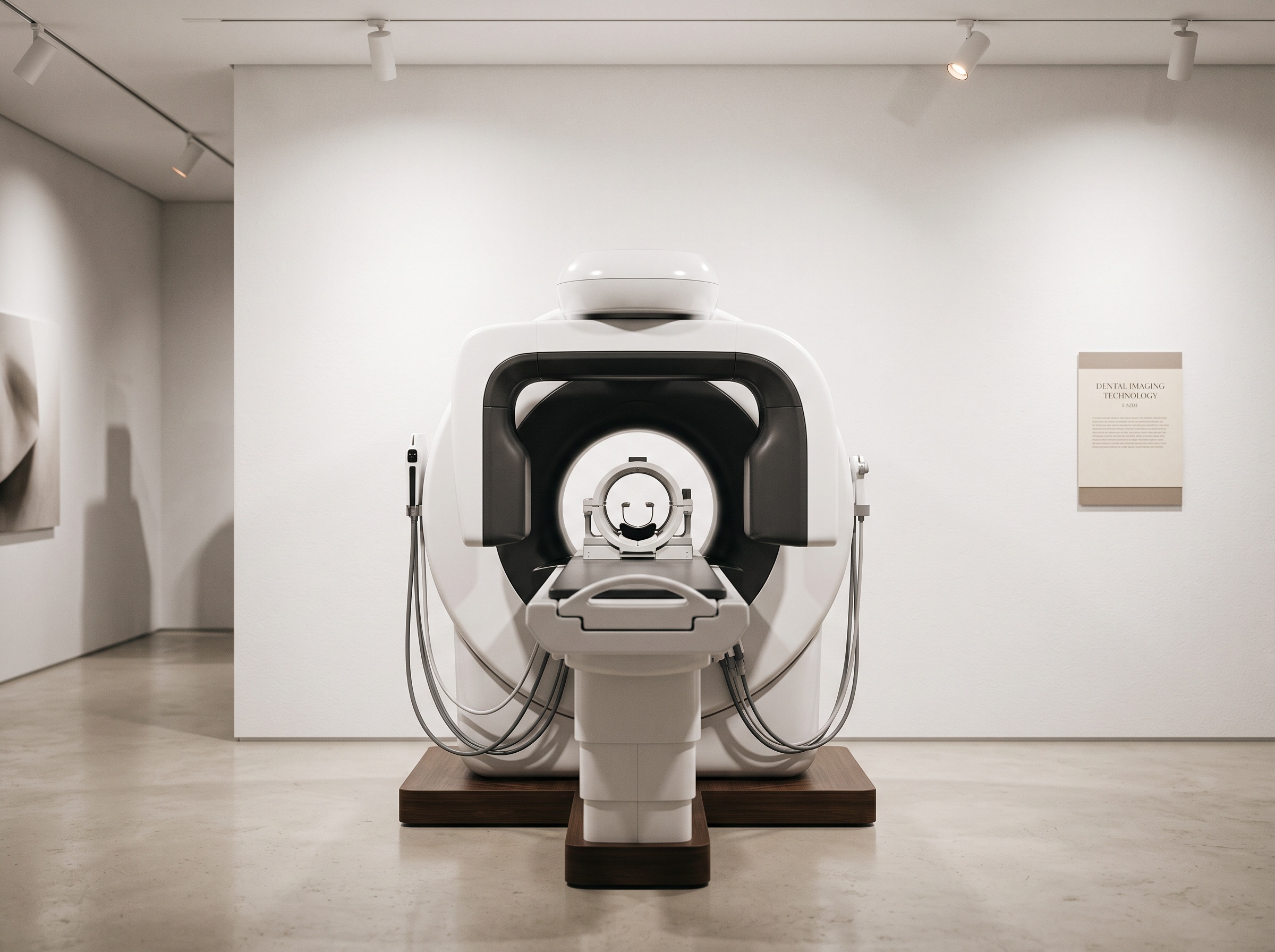
Honesty about an instrument includes honesty about its station. This first dental-dedicated MRI is not a chairside box for the solo practice; it is a scanner intended for hospitals, large dental clinics, and the academic centers that do dentomaxillofacial diagnostics, with a specialized dental coil and tailored software that bring an average workflow to under twenty minutes, brisk by MRI standards but still a considered procedure rather than a snapshot. Its natural referrals are the soft-tissue-rich problems that CBCT and plain film handle only partially: complex endodontic questions, periodontal and inflammatory assessment, the joint, the nerve, the orthodontic and surgical planning that benefits from seeing tissue as well as bone. For the everyday diagnostic question the familiar hierarchy still holds, the one we laid out in weighing the full-mouth series against cone-beam CT. MRI does not collapse that hierarchy; it adds a new rung at the top of it, reached when the diagnostic question is one that no amount of radiation was ever going to answer.
Future Developments
A cleared device at the referral centers is how nearly every imaging modality begins its life; the history of dental radiography itself is a long migration from the hospital and the specialist down to the operatory wall. It is reasonable to expect the dental MRI to travel the same road, toward smaller fields, lower-strength magnets friendly to a clinic’s floor plan, faster sequences, and eventually a form that a well-equipped practice can justify. As that happens, this gallery gains a subject it never truly had: the living soft tissue of the mouth, rendered in its own tonal language without a single photon of dose. The radiograph taught us to read bone as an image; the dental MRI asks us to learn to read the pulp, the ligament, and the disc the same way, as pictures with their own grammar of brightness and shadow. The instrument has cleared its first gate. The craft of reading it, patiently and well, is the work now beginning.
Sources & further reading:
- Dentsply Sirona Advances Dental Imaging With MRI – Dentistry Today
- First-ever Dental-Dedicated MRI Validated in Clinical Trial and Cleared by FDA for Advanced Soft-Tissue Diagnostics – Dentsply Sirona
No Comments