


February 7, 2026 The Geometry of an Honest Radiograph: Paralleling, the Bisecting Angle, and the Truth About Length
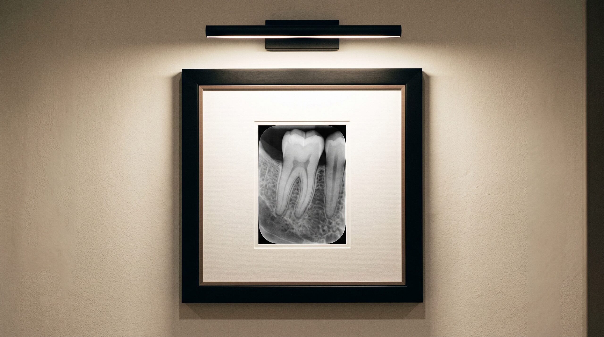
A dental radiograph arrives on the screen looking like a photograph, and that is its most persuasive lie. A photograph records light reflected from surfaces; a radiograph records a shadow — the pattern an X-ray beam leaves behind after passing through a tooth and striking a flat receptor. And a shadow, as anyone who has watched their own lengthen across a pavement at dusk knows, is an unreliable witness to size. It stretches and shrinks with the geometry of the light. The whole discipline of intraoral technique exists to keep that shadow honest: to make the image on the plate report the tooth’s true length, not a flattered or foreshortened version of it.
That honesty is not a matter of a better sensor or a sharper lens. It is a matter of arrangement — where the focal spot sits, how the receptor lies against the tooth, and the angle at which the beam is asked to travel. Get the geometry right and the radiograph becomes a faithful measurement. Get it wrong and it becomes a convincing forgery, dimensionally false in ways that no amount of contrast or resolution can undo.
Two Geometries, One Shadow
There are two classical ways to arrange the intraoral trinity of tooth, receptor, and beam, and they have contended with each other since the earliest decades of the field. In the bisecting-angle technique, the receptor is placed close against the tooth, resting at whatever angle the palate or floor of the mouth permits. Because tooth and receptor are not parallel, they form an angle between them; the operator imagines the line that bisects that angle and directs the central ray perpendicular to it. The reasoning is a neat piece of geometry — projecting a shadow perpendicular to the bisector makes the image length equal the true length, at least in theory.
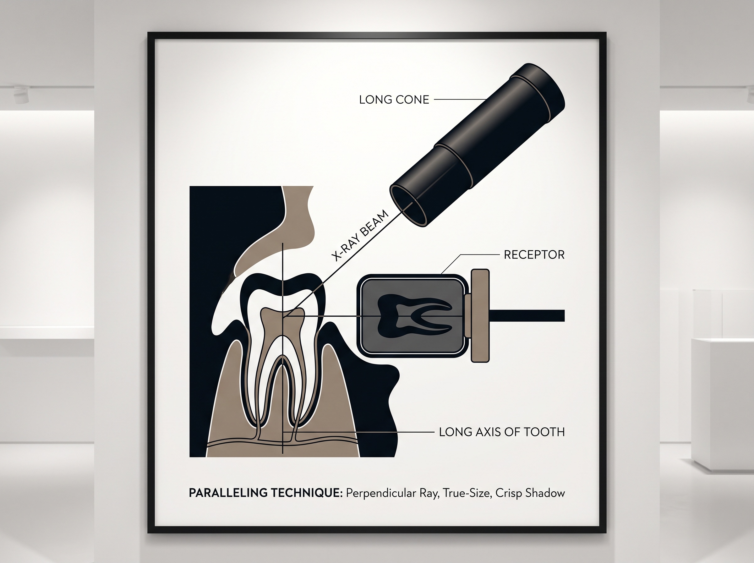
In the paralleling technique, the receptor is instead held away from the tooth, deeper in the mouth, positioned so that it lies parallel to the long axis of the tooth. The beam is then aimed at a right angle to both. There is no bisector to estimate, because there is no angle to bisect — the geometry is made simple and rigid by a holder that fixes receptor and beam in a fixed relationship. What the bisecting angle asks the operator to judge, the paralleling technique builds into a jig.
Foreshortening and Elongation: When the Image Lies About Length
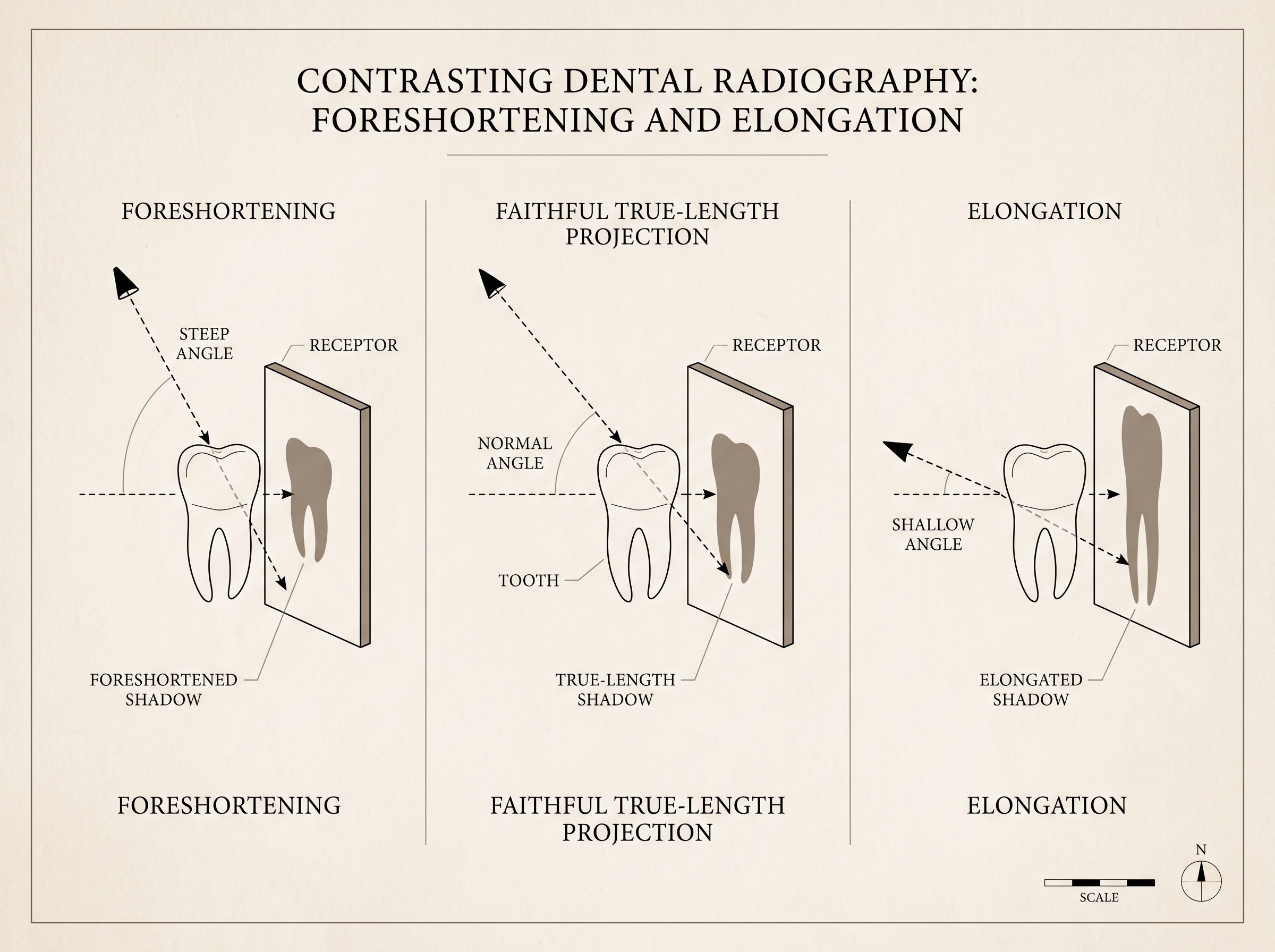
Both techniques are haunted by the same two ghosts, and their names describe exactly what they do. Foreshortening is the shadow made too short: it appears when the beam strikes the receptor at too steep a vertical angle, compressing the projected root into a stub that would flatter no clinician and mislead any measurement. Elongation is the opposite — a shadow drawn too long, produced when the vertical angle is too shallow, stretching the root toward a false apex.
The bisecting-angle technique lives on a knife’s edge between them. Its accuracy depends entirely on estimating the bisector correctly, and the human eye estimating an imaginary line inside a closed mouth is not a precision instrument. Aim a few degrees too steep and the tooth foreshortens; a few degrees too shallow and it elongates. Worse, because different structures sit at different angles, a single exposure can foreshorten the buccal roots while faithfully rendering the palatal one — distortion that varies across the very same film. The image is not uniformly wrong, which is more dangerous than if it were, because it looks internally plausible.
Why Paralleling Tells the Truer Story
The paralleling technique earns its status as the preferred method by removing the estimate. When the receptor lies parallel to the tooth and the beam meets both at ninety degrees, foreshortening and elongation have nowhere to enter — the projected length equals the true length because the geometry forbids the distortion rather than merely trying to cancel it. It is the difference between a level held against a wall and a carpenter squinting to guess the vertical.
But parallelism alone would introduce a different error. Holding the receptor away from the tooth means the tooth is no longer pressed against the plate, and any object held away from the receptor casts a magnified shadow, its penumbra softened and its edges blurred. The classic remedy is distance: a long cone — a longer source-to-object distance — narrows the beam so that the rays arriving at the tooth are very nearly parallel to one another. A near-parallel beam projects a near-true-size shadow with a crisp edge, even across the gap the paralleling technique requires. This is why the mature form of the method is properly called the paralleling, long-cone technique: parallelism handles the angle, and distance handles the magnification the parallelism would otherwise cost.
The Bisecting Angle’s Honest Case
None of this makes the bisecting angle worthless, and a refined clinician should know why it survives. Some mouths refuse the paralleling holder: a shallow palate, a strong gag reflex, an uncooperative floor of the mouth, or a child’s small arch can make it impossible to seat a receptor parallel to the tooth. In those moments the bisecting angle is not a compromise of laziness but of anatomy — a way to obtain a diagnostic image when the ideal geometry is physically denied. The skilled operator treats it as a fallback whose limitations are understood in advance: expect some dimensional infidelity, interpret length with caution, and never make a precise measurement — an implant length, a root canal working length — from an image the geometry cannot vouch for.
Reading Distortion in a Finished Image
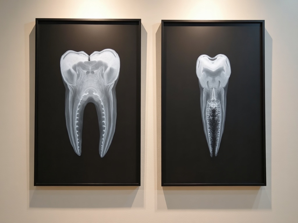
The craft does not end at exposure; it continues in interpretation. A trained eye reads a radiograph partly as a record of its own geometry. Roots that look improbably stubby against otherwise normal crowns whisper foreshortening. Roots stretched to implausible lengths, or a cusp tip projected far from where the anatomy says it should sit, betray elongation. When the same film shows one structure faithfully and its neighbour distorted, the reader knows the beam’s vertical angle was fighting the anatomy. To read these signatures is to recover the truth the geometry tried to hide — and to know when a measurement can be trusted and when the image must be retaken before anyone acts on it.
Future Developments
Three-dimensional imaging changes the terms of this old contest without ending it. Cone-beam computed tomography reconstructs a volume from many projections and, when properly calibrated, renders anatomy at true scale in every plane — an answer to the length problem that a single intraoral shadow can only approximate. Yet the intraoral radiograph endures for its resolution, its low dose, and its immediacy, and so its geometry still matters. The likeliest near future is not the retirement of the periapical but its guidance: receptor holders with alignment sensors, beam aiming assisted by the machine, and software that flags a foreshortened or elongated image before the clinician commits to it. The instinct is old — make the shadow tell the truth about length — and the tools grow quietly better at enforcing it. As always at PatientGallery, the finest image is not merely the sharpest but the most faithful, and fidelity, in radiography, begins with geometry.
Sources & further reading:
- Intraoral radiographic techniques: paralleling and bisecting-angle (Wikipedia, Dental radiography)
- White & Pharoah, Oral Radiology: Principles and Interpretation — projection geometry and image distortion
- Paralleling technique (Radiopaedia)
Sorry, the comment form is closed at this time.