


October 10, 2026 Mapping the Salivary Rivers: Sialography and the Craft of Imaging the Ducts That Keep the Mouth Alive
Almost everything dentistry images is solid. Enamel and dentin, cortical and trabecular bone, the hard geometry of a root and the engineered geometry of an implant — the discipline has spent a century learning to render stillness in exquisite detail. Yet one of the mouth’s most consequential structures is not solid at all. It is a plumbing system: three major salivary glands on each side, each draining through a slender duct into the oral cavity, carrying the fluid that lubricates speech, begins digestion, buffers acid, and keeps the tissues alive. When that plumbing blocks, narrows, or inflames, the patient knows it as a swollen, painful gland at mealtimes. To see why, you cannot photograph a surface or slice a bone. You have to picture a river. Sialography is the craft of doing exactly that.
Imaging a fluid, not a solid
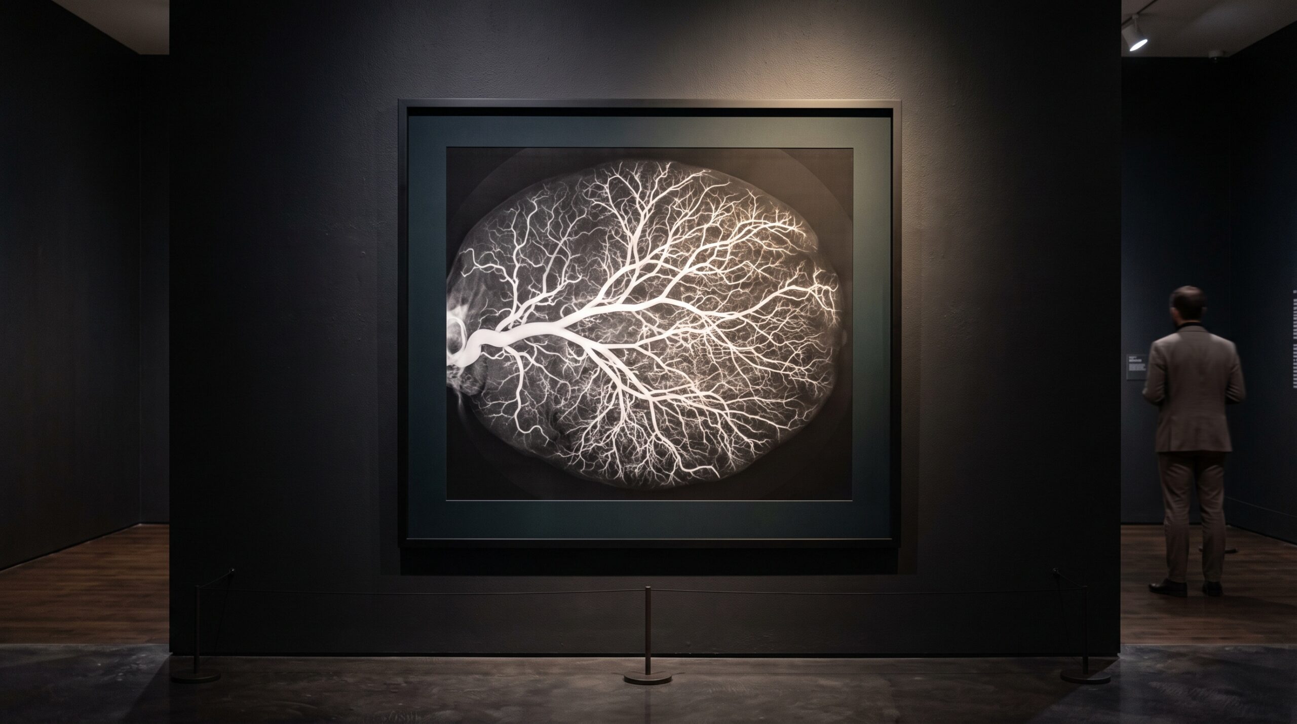
Sialography is, in its classic form, a radiographic technique in which radio-opaque contrast medium is instilled directly into a salivary duct and then imaged, so that conditions such as ductal obstruction, strictures, dilatations, stones, and salivary aplasia can be assessed. The logic is beautiful in its simplicity. A duct is a dark, soft-tissue channel that no plain radiograph can distinguish from everything around it. Fill that channel with something that stops X-rays, and the invisible becomes a bright, branching cast — the ductal tree drawn in light, from the single main trunk down through its finer and finer tributaries. The image is not of the gland but of the space inside it, and that negative space is where almost all benign salivary disease declares itself.
This is a different ambition from most of the imaging covered here. Where the bitewing reveals the hard-tissue story of caries and bone, a sialogram reveals a hydraulic one: patency, flow, the architecture of a conduit. It belongs to the same family of thought that treats the image as evidence rather than decoration, but its subject is motion arrested — a fluid system caught mid-flow.
The anatomy the technique exists to map
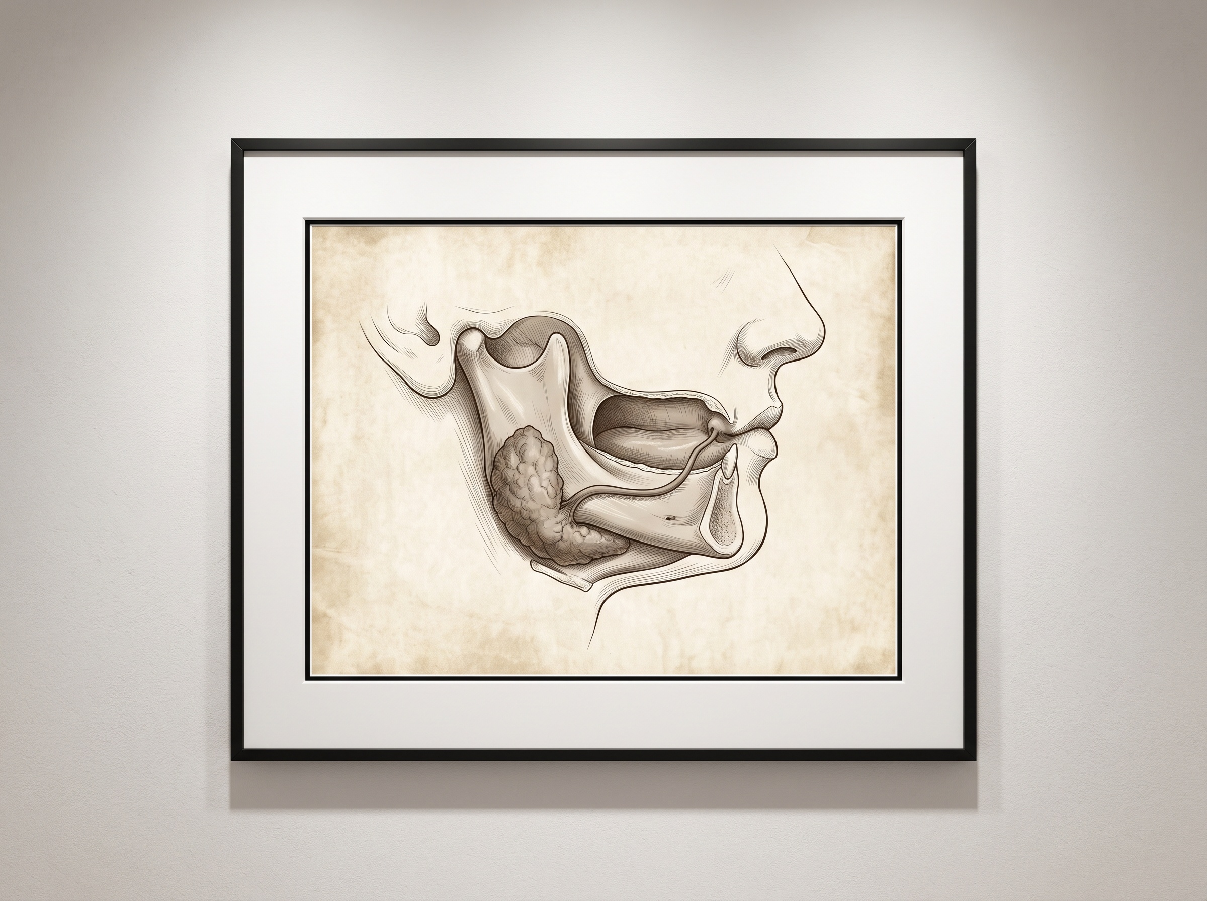
Three paired glands do the major work. The parotids, largest and set over the angle of the jaw, drain forward through a duct that opens inside the cheek opposite the upper molars. The submandibular glands sit beneath the floor of the mouth and drain upward and forward through a long duct to an opening under the tongue — an uphill path that, combined with their thicker secretion, makes them the usual home of salivary stones. The sublinguals are smaller and more diffuse. Each duct is a single slender lifeline, and sialography exists to trace that lifeline from its orifice to the deepest branch, mapping the salivary network and identifying the structural inconsistencies — obstructions, dilatations, and ductal variations — that a purely anatomical scan might never explain.
The craft of the conventional study
A conventional sialogram is a procedure of patience and delicacy. The duct orifice — a pinpoint on the cheek or beneath the tongue — is identified and gently dilated, and a fine blunt cannula is introduced. Contrast is instilled slowly, by hand, with the operator reading the patient’s sensation of fullness as a gauge; overfill and the medium extravasates, obscuring the very anatomy it was meant to reveal. Images are then made in a filling phase, when the tree is fully opacified and its branching pattern is read for strictures, dilatations, or the filling defect of a stone, and often again in an emptying phase, after a sialogogue such as a drop of lemon stimulates the gland, so that the clinician can judge how readily the duct clears — a functional reading layered onto the anatomical one.
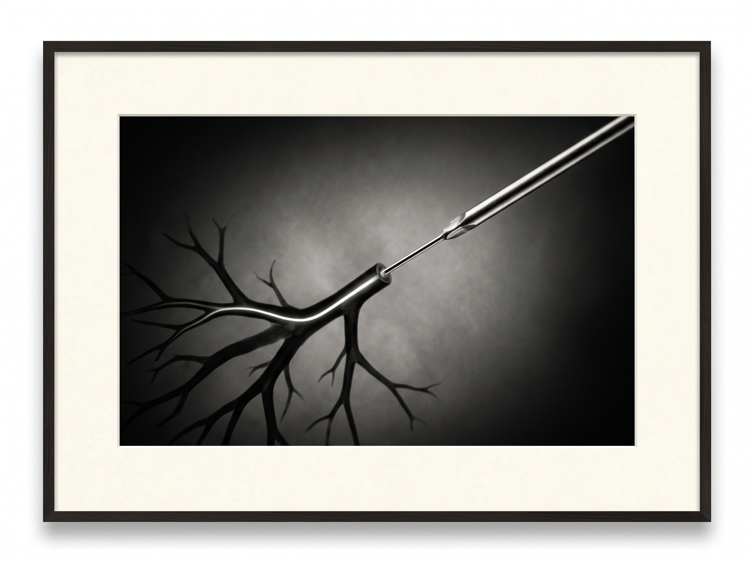
Every stage is a place where technique governs the result. The cannulation must not traumatize the orifice; the instillation must not perfuse too fast; the timing of the exposure must catch the tree at its most informative. It is, in the truest sense, an imaging craft — one in which the picture is only as good as the hand that filled the duct. Because the study uses both ionizing radiation and an injected contrast agent, it also carries the discipline that governs any dose decision in dentistry: the quiet obligation to justify and minimize every exposure, and clear contraindications in acute infection or known contrast allergy.
Saliva as its own contrast: the MR study
The most elegant modern turn is to dispense with the needle entirely. MR sialography exploits a simple fact: saliva is water, and water is bright on heavily T2-weighted, fat-suppressed MRI sequences. The fluid already sitting in the duct becomes its own intrinsic contrast, so the ductal tree can be visualized with no cannulation, no injected medium, and no radiation at all. For a patient with an acutely inflamed gland — precisely the case in which threading a cannula is most difficult and most contraindicated — this is transformative. Comparative work has found MR sialography a capable, non-invasive alternative for assessing salivary duct disease, trading a measure of fine ductal detail for the enormous advantage of leaving the gland untouched.
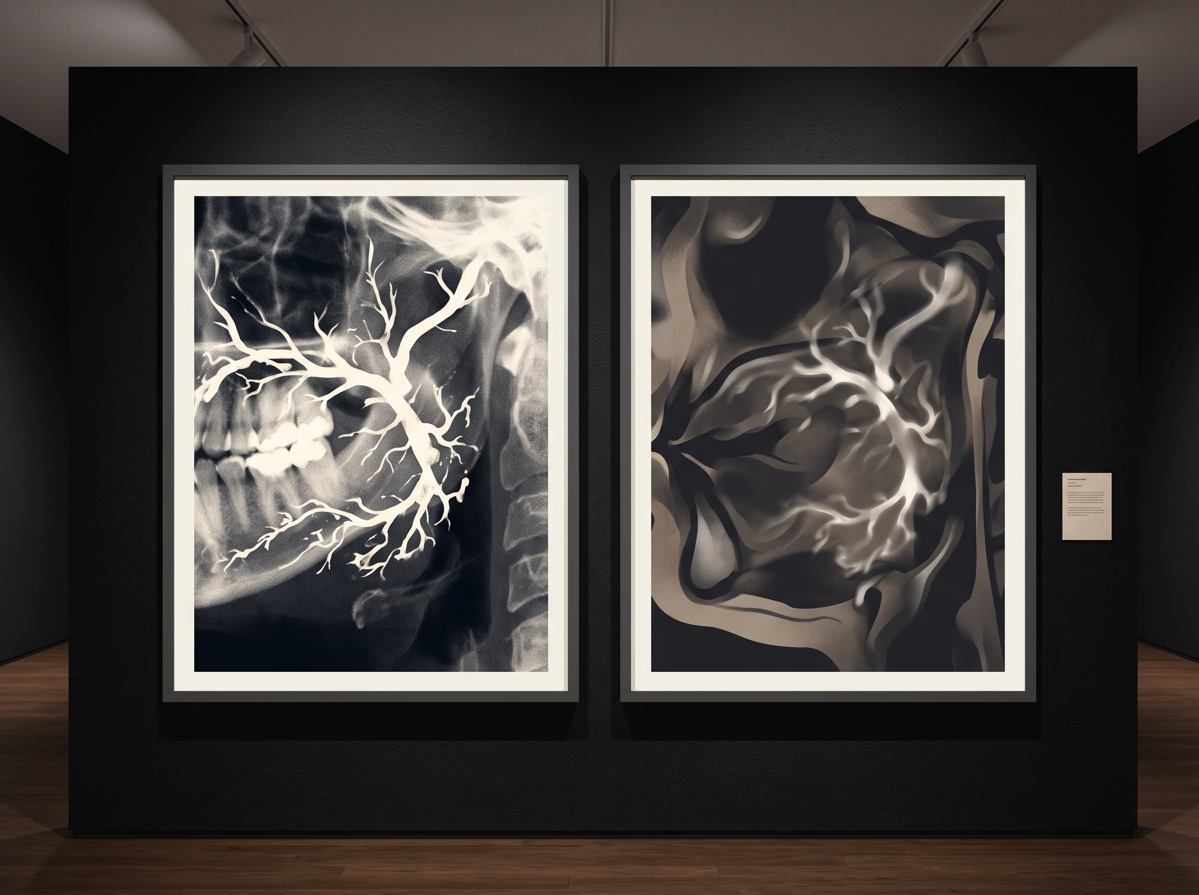
There is a conceptual kinship here worth savoring. Just as transillumination images a crack with light instead of radiation, MR sialography images a duct with the body’s own fluid instead of an injected dye — both are quiet refusals of the more invasive path when a gentler signal is already present. And because it is an MRI study, it inherits the same cross-sectional soft-tissue strengths that make MRI the reference for the temporomandibular joint, letting the reader see not only the duct but the gland parenchyma and surrounding tissue in the same examination.
Where it sits among the modern options
Sialography does not stand alone. Ultrasound is often the first look, cheap and radiation-free, able to spot a dilated duct or a shadowing stone in the superficial glands — and in ultrasound sialography, a duct can even be gently infused and watched in real time. Cone-beam and multidetector CT excel at dense stones and bony relationships. Sialendoscopy, the threading of a miniature endoscope into the duct, has blurred the line between seeing and treating, allowing stones to be retrieved and strictures dilated without sacrificing the gland. Against this crowded field, a 2026 case series makes the honest argument that conventional sialography still offers a unique advantage for mapping the ductal network and demonstrating the extent of benign obstructions and ductal variations — a whole-tree roadmap that the newer, more targeted tools do not always provide. The technique has not been replaced so much as given a sharper, narrower indication.
Future Developments
It is tempting to read a century-old injection study as a relic, and tempting is exactly the wrong instinct. Sialography’s future looks less like obsolescence and more like refinement: MR sialography maturing into a routine first-line map where availability allows, ultrasound-guided and endoscopic techniques merging diagnosis with therapy in a single sitting, and conventional contrast studies reserved for the questions only a full ductal roadmap can answer. The deeper lesson is one this gallery returns to often — that an imaging method earns its place not by novelty but by what it uniquely lets us see. The salivary ducts are rivers we will never look at directly. Whether we trace them with injected light, with the glow of their own fluid, or with sound, the craft is the same: to make a moving, hidden thing hold still long enough to be read, and to choose, each time, the gentlest means of seeing that still answers the question.
Sources & further reading:
- Clinical insights on sialography in the era of modern imaging: A case report of 5 cases (PubMed, 2026)
- Sialography — an overview (ScienceDirect Topics)
- Ultrasound Sialography / MR Sialography (Iowa Head and Neck Protocols, University of Iowa)
No Comments