


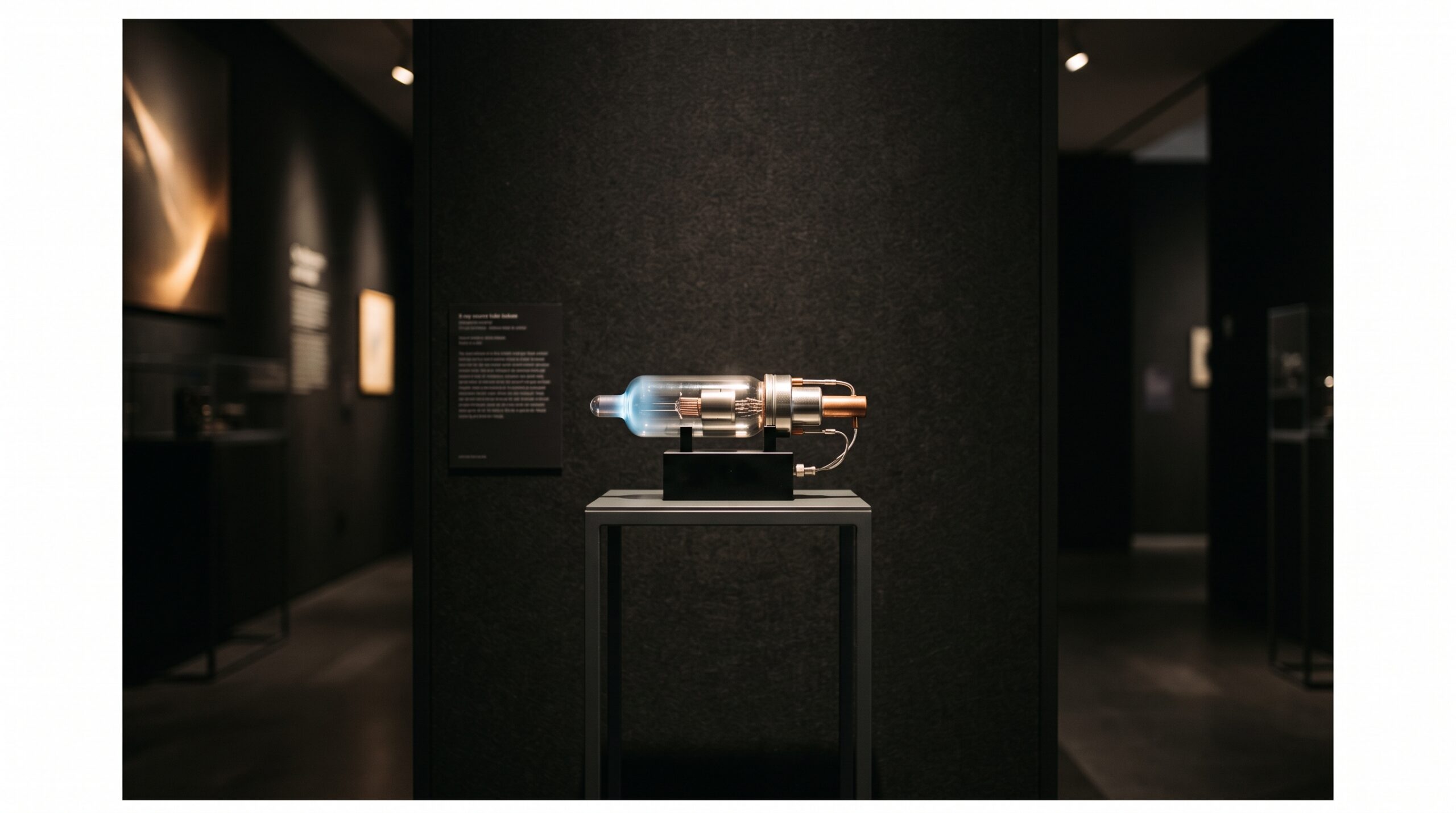
October 20, 2026 The Shrinking Source: Carbon-Nanotube X-Ray Tubes and the Quiet Reinvention of How a Dental Radiograph Begins
Almost everything written about dental imaging concerns the end of the chain. We admire the sensor and its shrinking pixels, the software that denoises and reconstructs, the screen on which the image is finally read. The beginning of the chain — the place the X-rays are actually made — we take for granted, because it has barely changed in a hundred years. Inside nearly every dental tubehead sits a design Coolidge would recognize from 1913: a wire filament heated white-hot until electrons boil off its surface, flung across a vacuum at a metal target, and converted, inefficiently and violently, into the beam that writes the picture. It works. It also wastes most of its energy as heat, cannot be switched on and off cleanly, and sets a hard floor under how small and how fast a source can be. A different way of releasing electrons — one that needs no heat at all — is now moving from the laboratory toward the operatory, and because it sits at the very start of the imaging chain, it quietly rewrites what is possible everywhere downstream.
The hundred-year-old flame
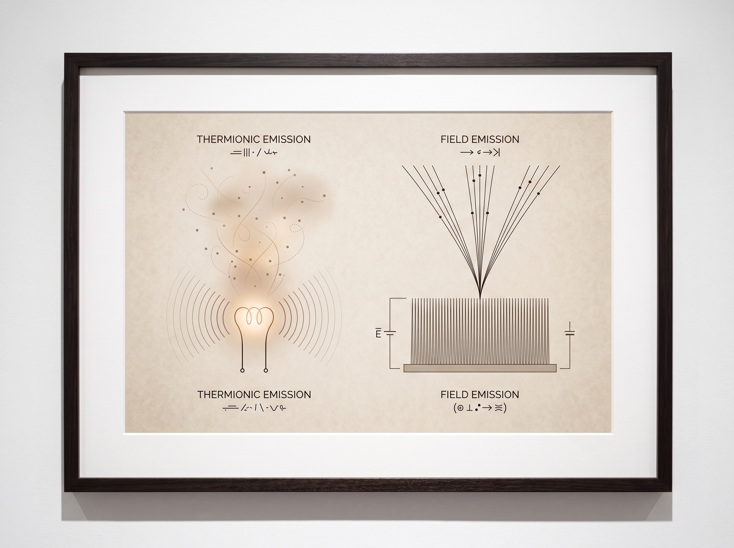
To appreciate the change, look honestly at the incumbent. Thermionic emission — the boiling-off of electrons from a hot filament — is a blunt instrument. The filament must be brought up to temperature before it will emit, so the source cannot turn on instantly or off cleanly; the exposure has a soft beginning and a soft end, a smear of radiation at each edge that contributes dose without contributing much image. The heat is relentless, which is why tubes need bulky anodes and cooling and why they age and sag and eventually fail. And the emitting surface is a glowing wire of finite size, which limits how small the effective focal spot can be — and the focal spot is the single most important determinant of how sharp a radiograph can ever be. Every blur budget in projection radiography starts here. The filament, in other words, is not a neutral given. It is a constraint we have simply learned to live with, the way one lives with a draft from an old window.
Electrons without heat
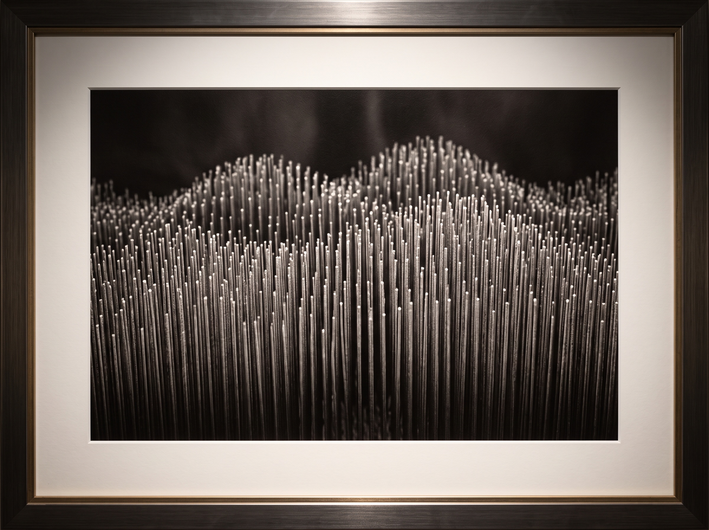
Field emission is the alternative, and it is an old idea finally given a good material. Instead of coaxing electrons out with heat, you pull them out with a powerful electric field — and the trick to doing that at sane voltages is geometry. An electric field concentrates fiercely at a sharp point, so if your emitter ends in a tip fine enough, the field at that tip becomes strong enough to tear electrons straight out of the metal, cold. The problem has always been finding something sharp enough, uniform enough, and durable enough to do this reliably across millions of tips at once. Carbon nanotubes turned out to be almost unfairly well-suited: they are essentially the sharpest stable objects chemistry can make, they conduct beautifully, and they can be grown in dense, aligned films — a carpet of microscopic lightning rods, each tip a point where electrons leap away the instant a field is applied. Drive that film with a voltage you can switch in nanoseconds and you have an electron source that is cold, instant, and controllable in ways the hot filament never was. The same physics that lets a mini X-ray source ride a dose budget down toward the floor is what makes NASA interested in it for medicine in orbit, where mass and heat and power are all scarce.
What changes when the source turns on like a lamp
Instant, clean switching is the first dividend, and it is larger than it sounds. A source that fires only during the precise window it is wanted — and stops dead the instant it is not — wastes no dose on the soft edges of an exposure, and it can be gated, synchronized, pulsed, and timed with an electronic precision a thermal filament can only approximate. Pulse it fast enough and you freeze motion, which matters for the squirming child and the trembling hand alike; the blur that a long exposure smears into a radiograph is partly a blur of time, and a source that can strobe removes it. Run it cold and the focal spot can be made genuinely tiny and stay that way, which lifts the ceiling on resolution rather than chasing it only at the sensor. And without the heat and the heft, the whole source shrinks — which is why the most visible beachhead for this technology so far is the handheld intraoral unit, light enough to carry, that increasingly relies on these cold emitters rather than a miniature hot tube. The same cleanliness that enables fast switching also pairs naturally with techniques that demand many near-identical exposures, like the subtraction methods that reveal change over time only when each image is made the same way as the last.
From one source to many
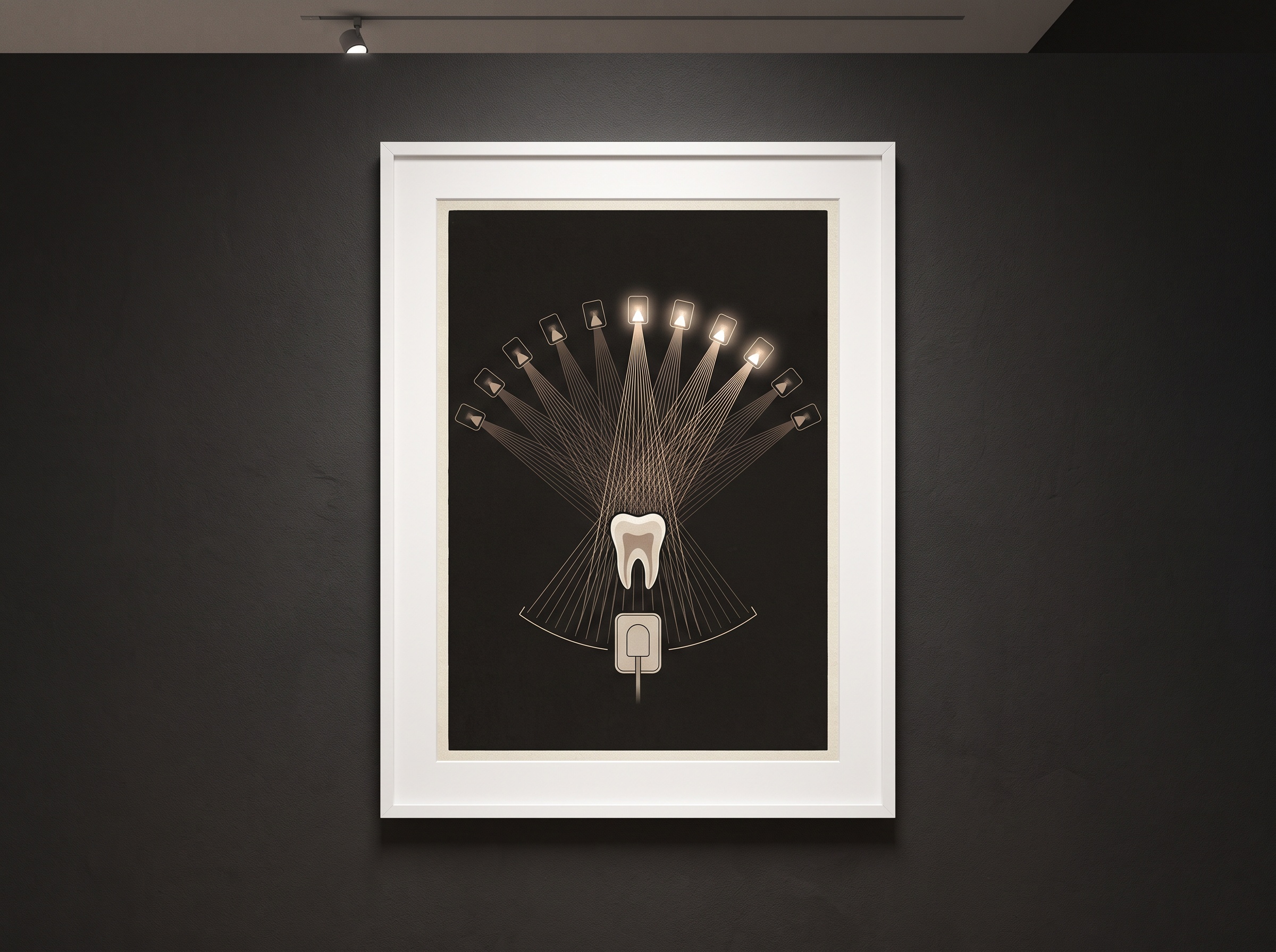
The deeper consequence is architectural, and it is the one that excites the people building these systems. Because a nanotube emitter is a flat film addressed by electronics rather than a single hot wire, you are no longer limited to one source. You can lay down many of them, each its own tiny tube, and fire them individually — a distributed, electronically multiplexed array of X-ray sources where there used to be exactly one. Arrange a row of them in an arc and fire them in sequence, and you capture a subject from many slightly different angles without moving anything at all. That is the principle behind stationary intraoral tomosynthesis: depth information, a stack of thin focal planes teased out of a stationary sensor and a stationary bank of sources, with none of the mechanical complexity of a rotating gantry. It is a genuinely new kind of dental image — more than a flat radiograph, less than a full CBCT volume, and physically possible only because the source stopped being a single object. The fast, independent switching also opens a door onto spectral imaging, where rapidly alternating beam energies let the machine begin to tell materials apart rather than merely map their density.
The honest ledger
None of this has displaced the hot filament yet, and it is worth saying why. The total X-ray output a nanotube cathode can sustain is still, for many sources, below what a thermionic tube delivers, which constrains the techniques it can serve; the emitter films can degrade, poison, or arc, and keeping emission uniform across a large array over years of clinical use is a real materials-engineering burden, not a solved problem. The high vacuum these devices demand is unforgiving of contamination. These are the frictions of a technology crossing from the bench into the operatory — the same awkward adolescence every imaging advance passes through — and they are reasons the transition is arriving first where the advantages are decisive and the demands modest: the portable unit, the specialized tomosynthesis head, the research bench. The filament will not vanish overnight. But the direction of travel is not in doubt.
Future Developments
The horizon here is a fully pixelated X-ray source: a flat panel of individually addressable nanotube emitters facing a flat panel of detectors, the beam no longer a single cone from a single point but a programmable field that can be shaped, steered, and sculpted in software — the emitting side of imaging finally becoming as digital and as flexible as the receiving side has been for twenty years. Pair that with the reconstruction mathematics already reshaping how we build volumes from projections and the low-dose learning methods that recover clean images from sparse data, and you can imagine dental radiography that chooses, exposure by exposure, exactly which rays to send and no others — all the information, none of the waste. For a gallery that treats the image as the art, there is a fitting lesson in looking this far upstream. The picture we frame on the wall is only ever as good as the light that made it, and for a century that light has come from a glowing wire we stopped noticing. Reinvent the source, and you reinvent the image — quietly, at the very place it begins.
No Comments