Logo Logo Logo Logo Logo
  • Home
  • Download
  • About
  • Blog
  • Home
  • Download
  • About
  • Blog
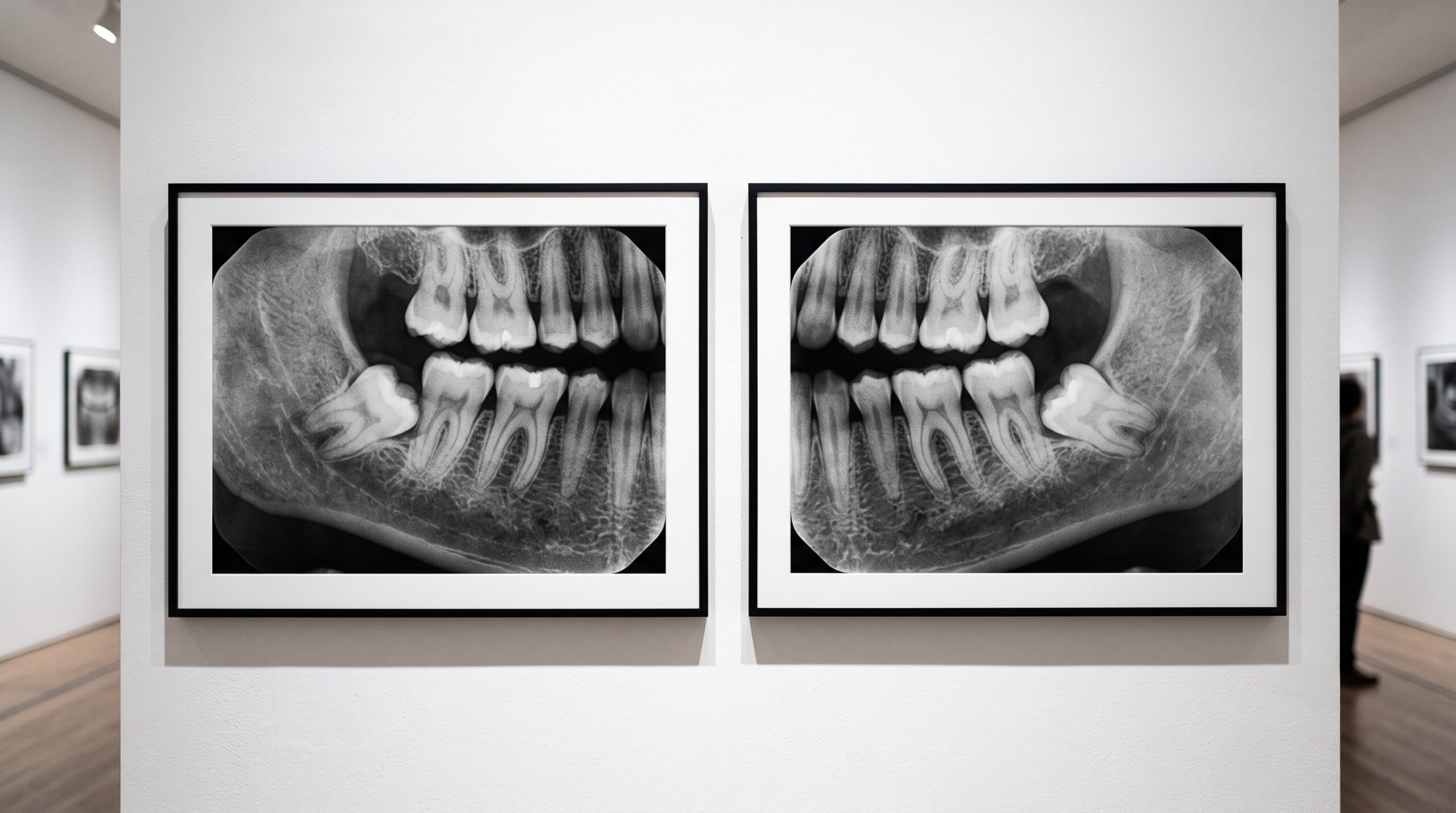
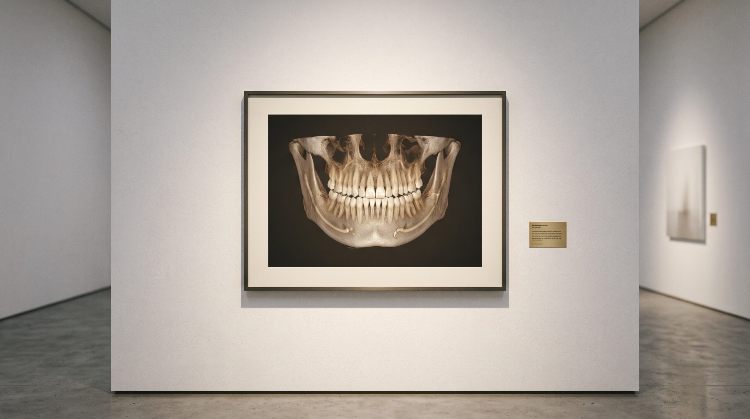
A framed panoramic dental radiograph on a gallery wall with a faint translucent ghost shadow floating over the opposite side of the jaw

22 Aug The Ghost on the Other Side: How a Panoramic X-Ray Paints a Shadow Where No Bone Exists

Posted at 11:31h in Dental Imaging by Raymond 0 Comments
0 Likes

There is a second image hiding inside every panoramic radiograph: faint, doubled, floating on the wrong side of the jaw. It is not a machine fault. It is geometry - and learning to read it is a quiet mark of the craft....

Read More
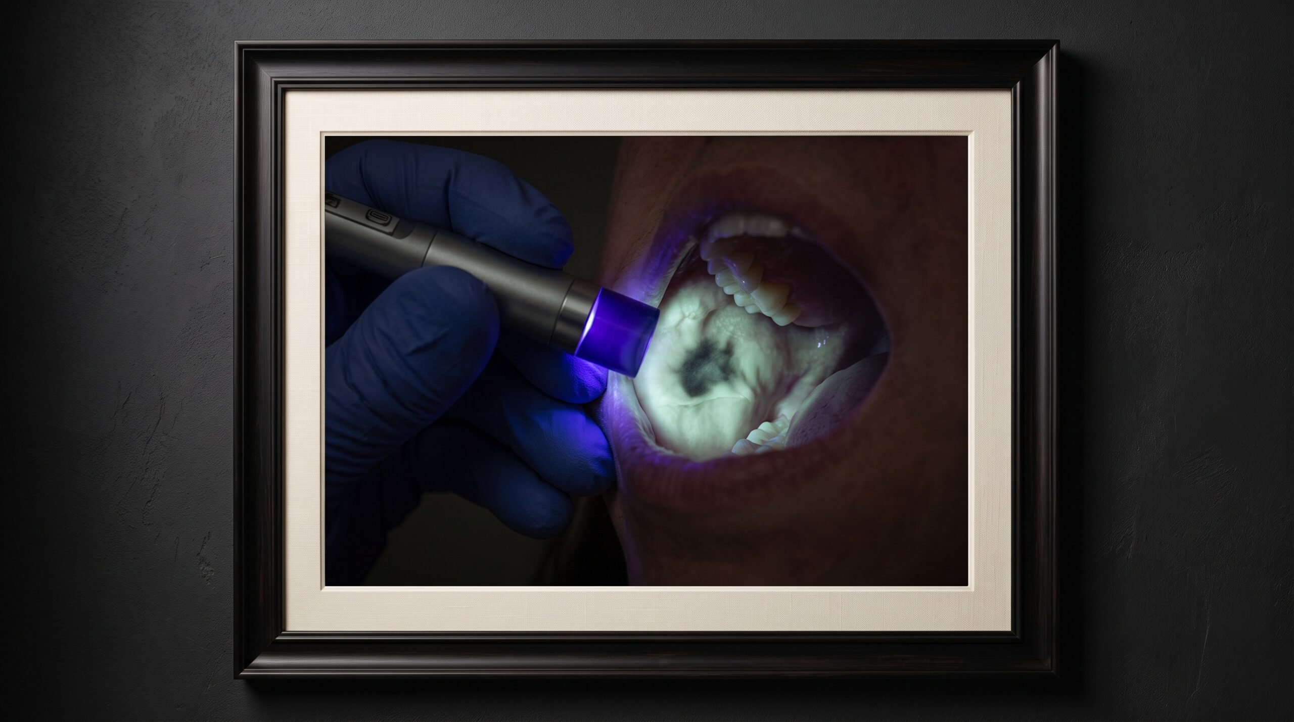
Two framed images of the same molar side by side - a color surface scan and a grey radiograph - with light suggesting they are merging

21 Aug Two Ways of Seeing, One Frame: Fusing the Intraoral Scan and the Radiograph Into a Single Image

Posted at 10:38h in Dental Imaging by Raymond 0 Comments
0 Likes

One instrument captures the surface and refuses to look beneath it. The other looks straight through and forgets the surface exists. For decades the clinician fused them in the mind. Now the software does it - and the craft is changing....

Read More
A framed dental radiograph on a gallery wall with a small brass placard beneath it bearing a single glowing number

20 Aug The Dose You Cannot See: Exposure Index, Dose Creep, and the Number a Forgiving Sensor Hides

Posted at 11:39h in Dental Imaging by Raymond 0 Comments
0 Likes

Film punished overexposure by going dark. Digital rewards it with a cleaner picture - which is exactly how dose creeps upward, invisibly. Here is the number that fixes it....

Read More
A framed dental radiograph with a soft translucent grey veil drifting over its lower half, washing out the contrast there

19 Aug The Scattered Photon: How Compton Scatter, Collimation, and the Missing Grid Shape Contrast in Intraoral Radiography

Posted at 10:37h in Dental Imaging by Raymond 0 Comments
0 Likes

A radiograph's contrast is built by the photons that are cleanly absorbed and the ones that pass straight through. The scattered photon belongs to neither, and it is the quiet vandal of the image - a fog with no source that dental imaging fights not...

Read More
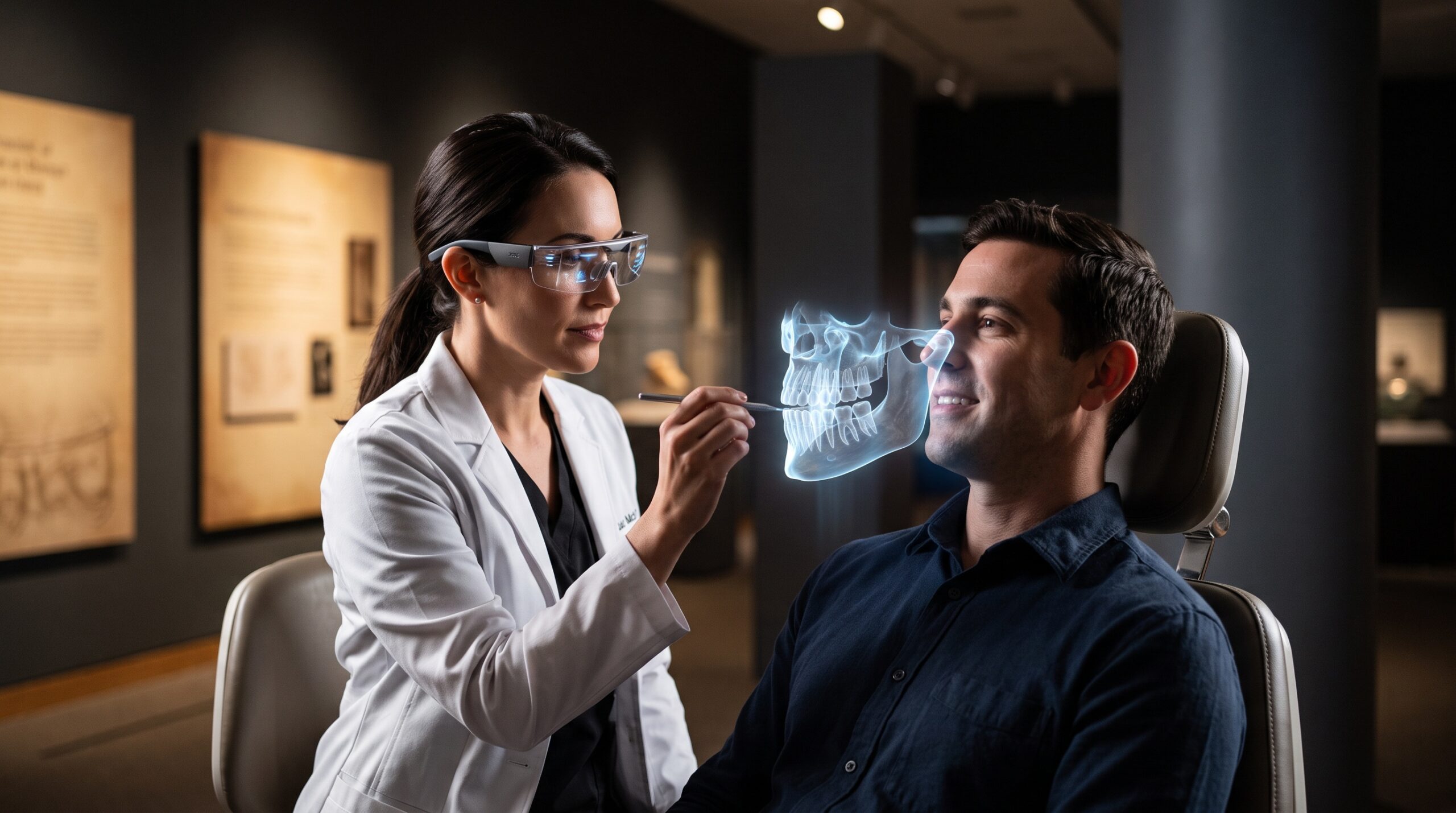
A framed dental radiograph on a gallery wall with a small blank museum placard beside it under refined lighting

18 Aug The Placard Beside the Picture: How Structured Reporting Makes a Dental Radiograph Legible

Posted at 10:38h in Dental Imaging by Raymond 0 Comments
0 Likes

In a gallery, the placard beside the artwork decides how much of the work you actually take in. The radiograph has a placard too - the report - and a new comparative study shows that giving it a fixed structure, rather than free-flowing prose, measurably...

Read More
A diagnostic display monitor shown as framed gallery art, its screen holding a dental radiograph in a smooth ramp of grays

17 Aug The Last Instrument: Diagnostic Display Calibration and the DICOM Grayscale Standard for Reading a Radiograph

Posted at 11:38h in Dental Imaging by Raymond 0 Comments
0 Likes

Every radiograph ends its journey on a screen, and the screen is the one instrument in the imaging chain that most practices never calibrate. The DICOM grayscale standard exists because the human eye reads brightness unevenly — and because a monitor that does not account...

Read More
A framed dental radiograph under gallery lighting showing a soft diffuse dark band at the neck of a tooth between crown and bone

16 Aug Cervical Burnout: The Radiolucent Illusion at the Tooth’s Neck That Mimics Caries

Posted at 13:39h in Dental Imaging by Raymond 0 Comments
0 Likes

Between the bright cap of the enamel and the pale crest of the bone runs a soft dark band along the neck of many teeth. It can look, unmistakably, like decay eating into the root. Often it is nothing of the kind — it is...

Read More
A thin scintillator screen framed as gallery art, faintly glowing with soft internal light

15 Aug The Needle Forest: How Columnar Cesium Iodide and the Scintillator Decide What a Sensor Can See

Posted at 10:37h in Dental Imaging by Raymond 0 Comments
0 Likes

A digital dental sensor does not, in most cases, feel the X-ray at all. It watches a thin crystalline screen flash with light and reads that instead. The material of that screen — whether a forest of light-piping cesium iodide needles or a settled bed...

Read More
A framed dental radiograph under gallery lighting showing a smooth density gradient from one edge to the other

14 Aug The Anode Heel Effect: The Invisible Gradient Shading Every Radiograph

Posted at 10:38h in Dental Imaging by Raymond 0 Comments
0 Likes

Every X-ray beam leaves the tube already uneven — brighter toward the cathode, dimmer toward the anode. The anode heel effect is the quiet gradient built into the physics of the source itself, and it shades the density of an image before the anatomy is...

Read More
A framed dental radiograph split into a soft honest half and a hyper-processed half with bright edge halos, under gallery lighting

13 Aug The Seductive Edge: How Sharpening, Contrast, and Noise Reduction Can Deceive on a Digital Dental Radiograph

Posted at 10:36h in Dental Imaging by Raymond 0 Comments
0 Likes

The radiograph a clinician reads is not the image the sensor captured. Between the photons and the screen lies a processing pipeline that sharpens, stretches, and smooths — and each of those kindnesses can quietly lie....

Read More
  • 1
  • 2
  • 3
  • 4
  • 5
  • 6
  • 7
  • 8
  • 9
  • 10
  • 11
© Copyright 2020, Raster Builders Inc. All Rights Reserved.