Logo Logo Logo Logo Logo
  • Home
  • Download
  • About
  • Blog
  • Home
  • Download
  • About
  • Blog
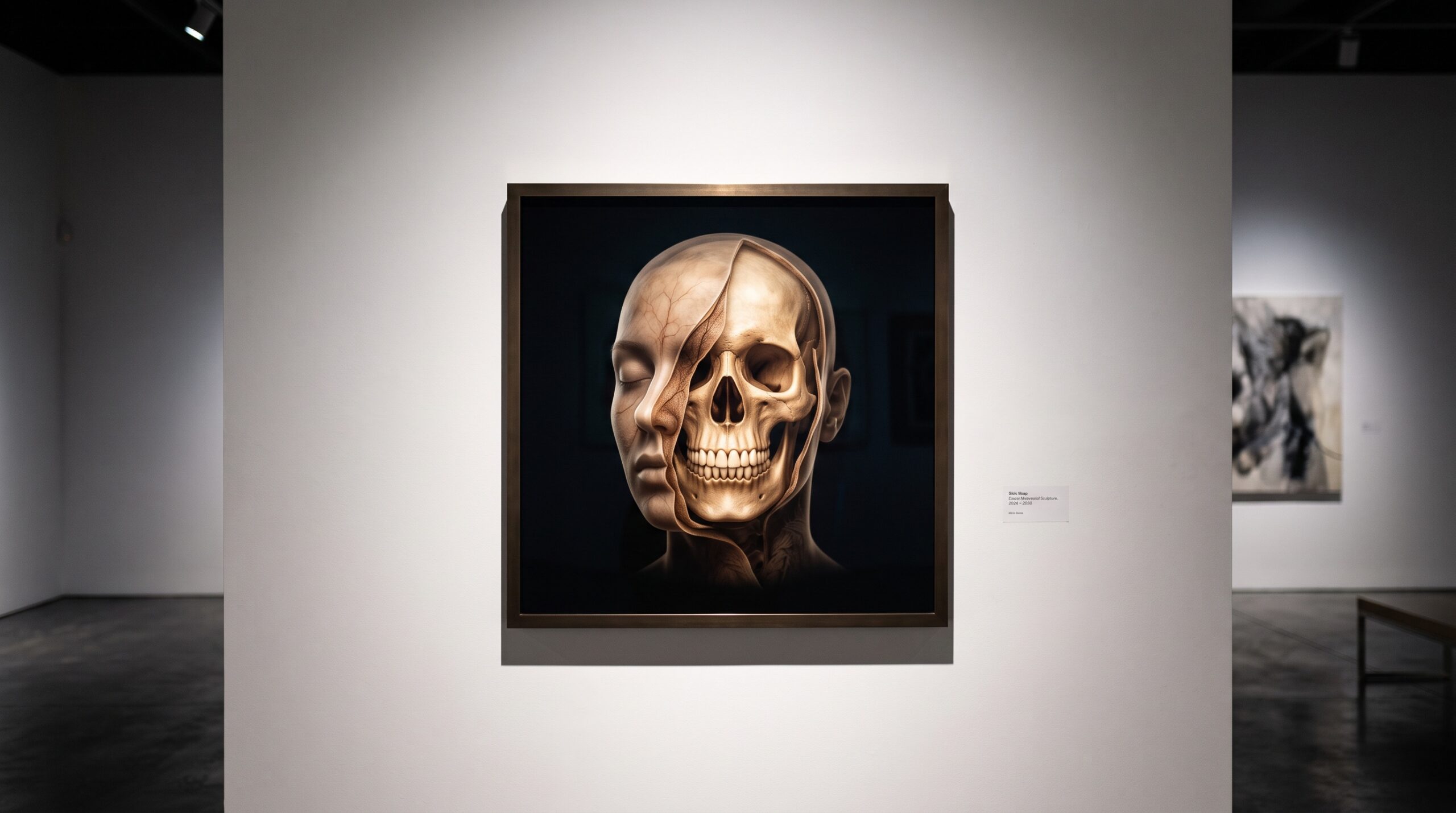
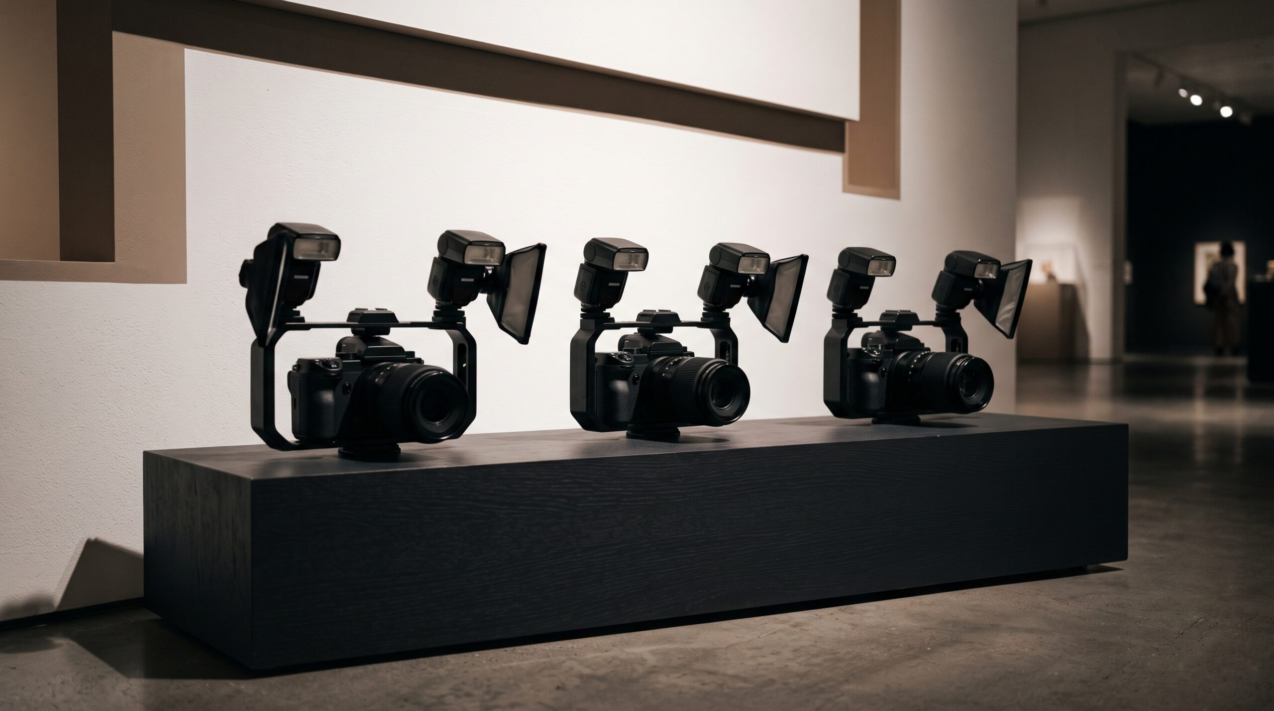
A compact handheld portable X-ray device shown as luminous framed art against a gallery white wall under museum lighting

17 Sep The X-Ray That Fits in a Bag: NASA’s Portable Imaging Trials and the Craft of Radiography Far From Any Hospital

Posted at 10:38h in Dental Imaging by Raymond 0 Comments
0 Likes

A broken bone or a cracked tooth a quarter-million miles from the nearest hospital is a problem of imaging before it is a problem of medicine. To answer it, NASA's Glenn Research Center has been testing portable, handheld X-ray systems built to work in a...

Read More
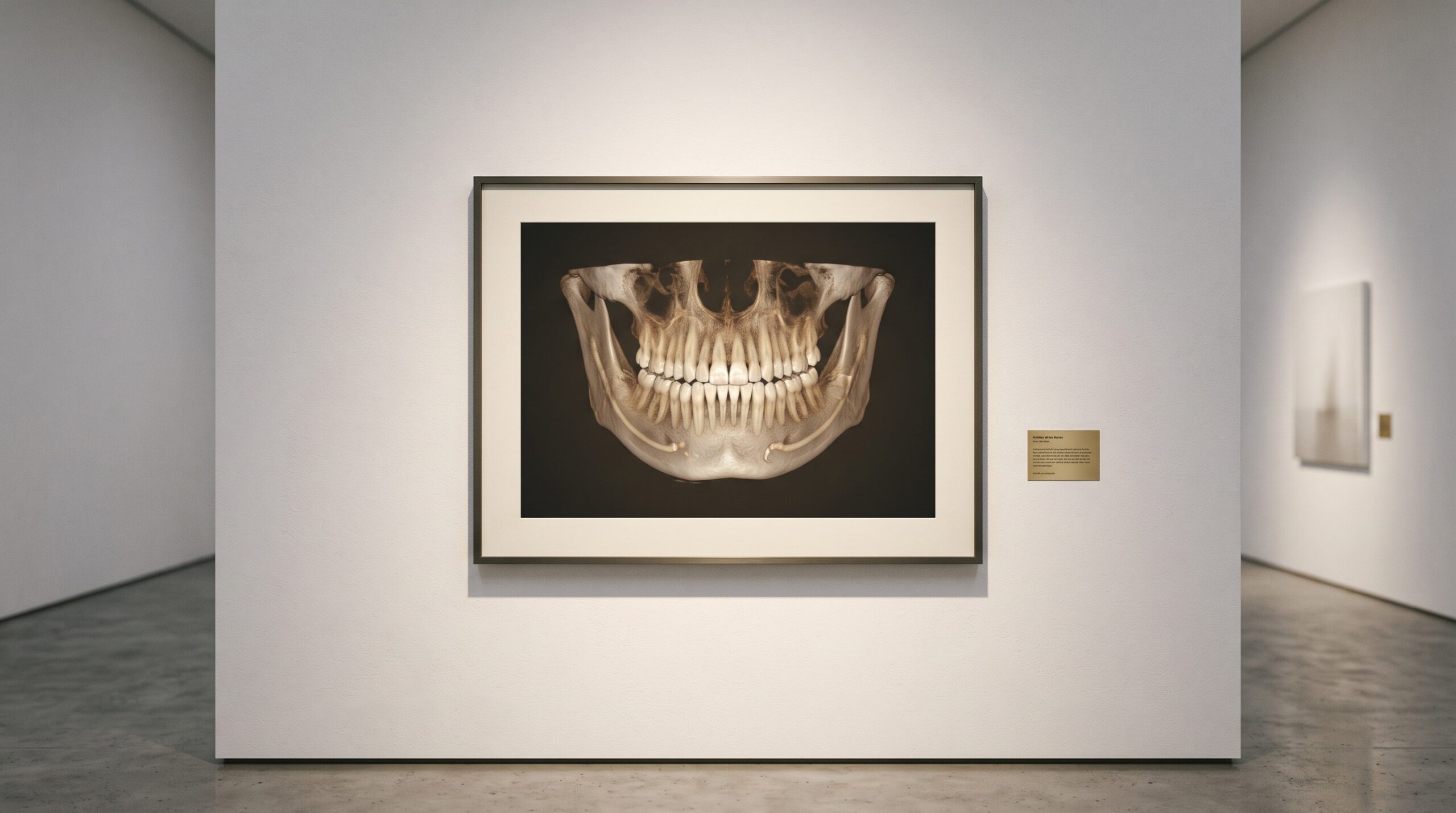
A panoramic dental radiograph shown as framed art, the sweeping jaw glowing, with the bright dense ribbon of cortical bone along the lower mandibular border drawn out as the subject

16 Sep The Bone at the Bottom of the Frame: How a Panoramic X-Ray Quietly Screens for Osteoporosis

Posted at 10:40h in Dental Imaging by Raymond 0 Comments
0 Likes

A panoramic radiograph is ordered to survey the teeth, the jaws, the developing third molars - and along the lower border of the mandible it records something no one asked it to. The inferior cortex of the jaw, that bright ribbon of dense bone at...

Read More
A translucent three-dimensional dental volume shown twice as one form split by an invisible seam into two subtly different luminous readings, one warm and one cool

15 Sep Two Energies, One Scan: How Spectral CBCT Tells Bone From Metal

Posted at 13:12h in Dental Imaging by Raymond 0 Comments
0 Likes

A conventional cone-beam scan measures one thing: how strongly each speck of tissue absorbs an X-ray beam. But absorption alone is ambiguous - a sliver of dense enamel and a whisper of metal can cast the very same grey. Spectral, dual-energy CBCT reads two different...

Read More
A translucent molar seen in three dimensions with its branching internal canal system glowing faintly inside, a delicate extra canal becoming visible behind a larger one

14 Sep The Hidden Architecture of a Tooth: What Cone-Beam CT Reveals That a Flat Radiograph Cannot

Posted at 10:37h in Dental Imaging by Raymond 0 Comments
0 Likes

For a century the radiograph told us almost everything about a tooth - and quietly kept one secret. Pressing a three-dimensional root system flat onto a single plane, it overlapped canals, hid the ones tucked directly behind others, and turned a fused, ribbon-like root into...

Read More
A single translucent tooth shown as a colour surface shell perfectly overlaid on a faint glowing interior of root and structure, aligned as one object

13 Sep One Mouth, Two Truths: The Craft of Fusing an Intraoral Scan With a Radiograph

Posted at 10:37h in Dental Imaging by Raymond 0 Comments
0 Likes

The intraoral scanner records the mouth as it looks - a flawless colour surface, and nothing beneath it. The radiograph records the mouth as light passes through it - the roots, the bone, the hidden lesion - and almost nothing of the surface. Each is...

Read More
A luminous tooth casting a soft graduated shadow with a fading penumbra edge, lit by a small glowing rectangular source above

12 Sep The Blur Built Into Every X-Ray: Focal Spot Size, Penumbra, and the Geometry of a Sharp Radiograph

Posted at 10:38h in Dental Imaging by Raymond 0 Comments
0 Likes

Every dental radiograph carries a blur that no detector, no software, and no amount of retaking can fully remove - because the X-ray source is not a point of light but a small glowing area on a metal target, and every point on that area...

Read More
A luminous colour 3D digital model of a child's dental arch displayed as gallery art, one tooth showing a faint chalky white lesion

11 Sep The Mouth on the Screen: Diagnosing Childhood Decay From a 3D Intraoral Scan

Posted at 10:39h in Dental Imaging by Raymond 0 Comments
0 Likes

For a century, diagnosing decay in a small child's mouth has meant one thing: a clinician, a mirror, a light, and a cooperative patient sitting still long enough to be examined. A 2025 Australian study quietly proposed an alternative. Capture a colour three-dimensional scan of...

Read More
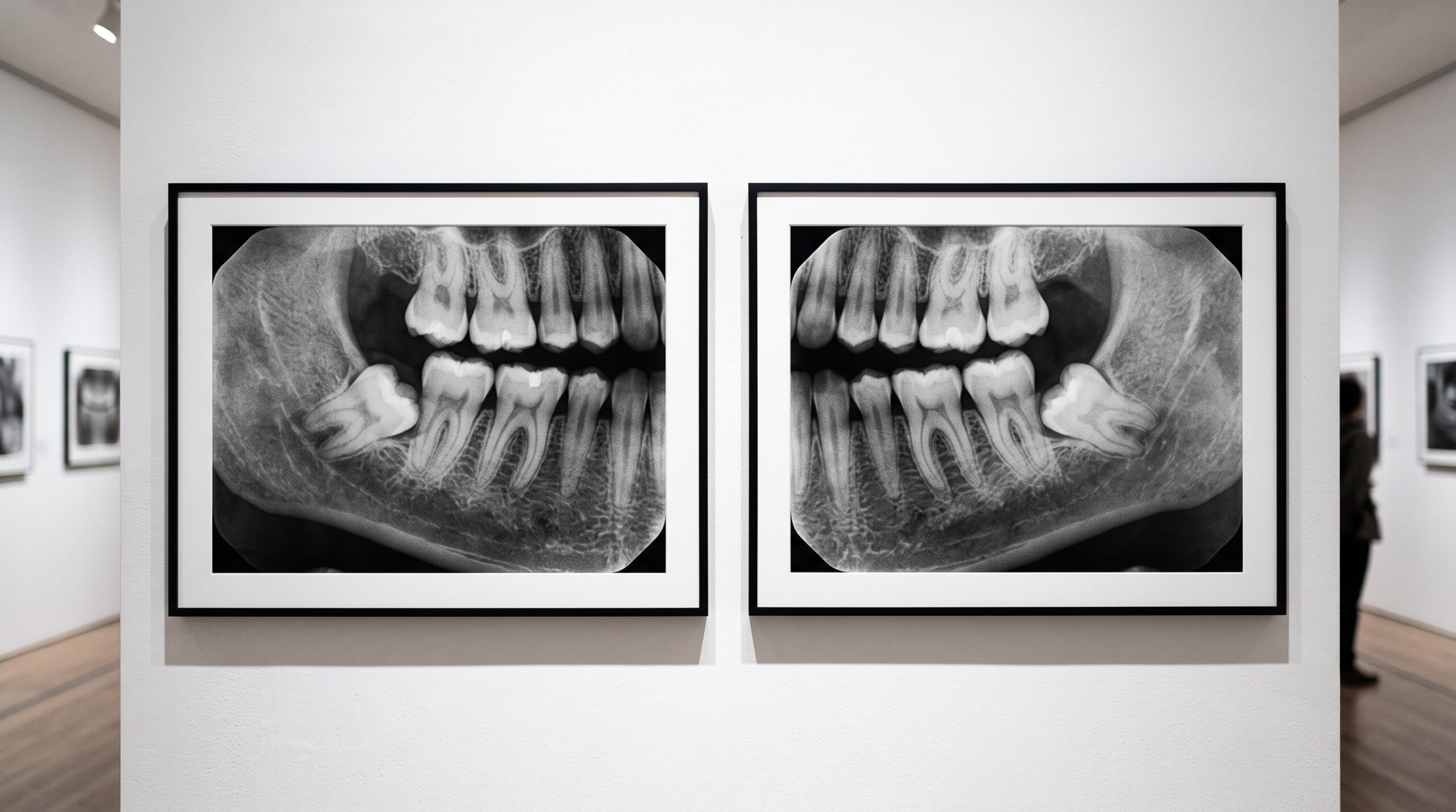
A gallery triptych: two near-identical bone radiographs and a third flat-grey panel showing only the glowing region where they differ

10 Sep The Art of Seeing Change: Digital Subtraction Radiography and Imaging What the Eye Cannot

Posted at 11:30h in Dental Imaging by Raymond 0 Comments
0 Likes

Two radiographs of the same tooth, taken months apart, can look identical to a trained eye while a real lesion quietly advances between them. Digital subtraction radiography is the technique that makes that invisible change visible: align the two images perfectly, subtract one from the...

Read More
Two framed panels in a gallery - an open door of light and a closed gate - with a radiograph between them

10 Sep Open API vs Partner-Gated: Which Dental AI Vendors You Can Actually Integrate With

Posted at 09:46h in Dental Technology by Raymond 0 Comments
0 Likes

A dental AI tool is only as useful as the door it will open for you. Some vendors publish a documented API and speak DICOM fluently; others hide behind an approved-partner gate you cannot unlock yourself. For anyone whose imaging software holds the radiographs, that...

Read More
A single empty ornate frame on a gallery wall, evoking the radiograph deliberately not taken

09 Sep The Discipline of Restraint: The New Imaging Guidance and the Art of the Radiograph Not Taken

Posted at 10:39h in Dental Imaging by Raymond 0 Comments
0 Likes

The most revealing decision a clinician makes about a radiograph is often whether to take it at all. The first ADA and AAOMR patient-selection recommendations in more than a decade - and the first to address cone-beam CT - reframe imaging not as a schedule...

Read More
  • 1
  • 2
  • 3
  • 4
  • 5
  • 6
  • 7
  • 8
  • 9
  • 10
  • 11
  • 12
  • 13
  • 14
  • 15
© Copyright 2020, Raster Builders Inc. All Rights Reserved.