Logo Logo Logo Logo Logo
  • Home
  • Download
  • About
  • Blog
  • Home
  • Download
  • About
  • Blog
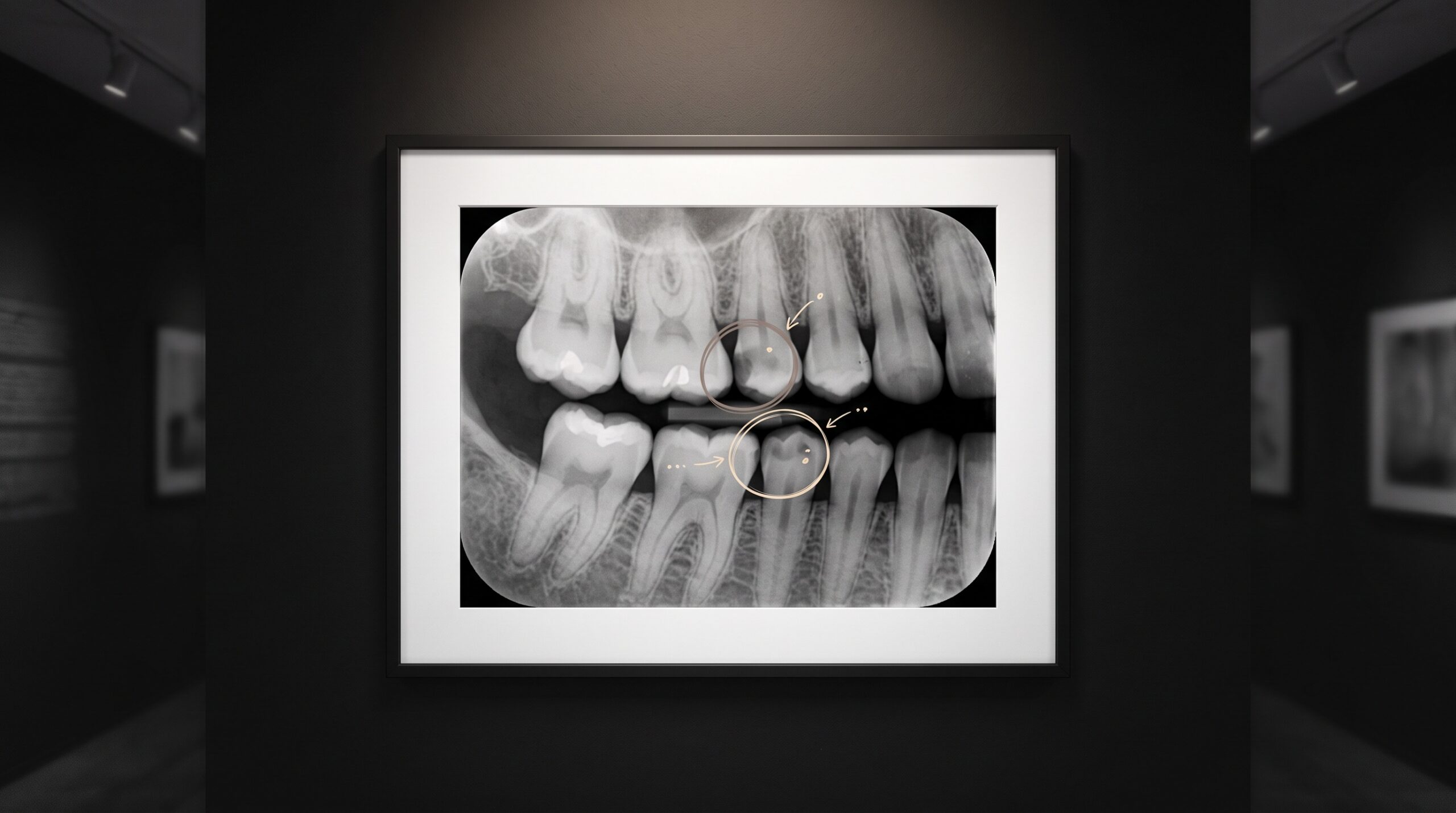
A framed dental bitewing radiograph on a gallery wall with faint glowing rings marking two suspected findings between the teeth

25 Aug Pearl on the Radiograph: FDA-Cleared Detection and How the Image Gets There

Posted at 09:44h in Dental Imaging by Raymond 0 Comments
0 Likes

Pearl's Second Opinion is the most-cleared name in dental AI - intraoral, panoramic, and now CBCT. The detections are the spectacle; the plumbing that carries the image out of the operatory and back is the part that actually decides who can use it....
Read More
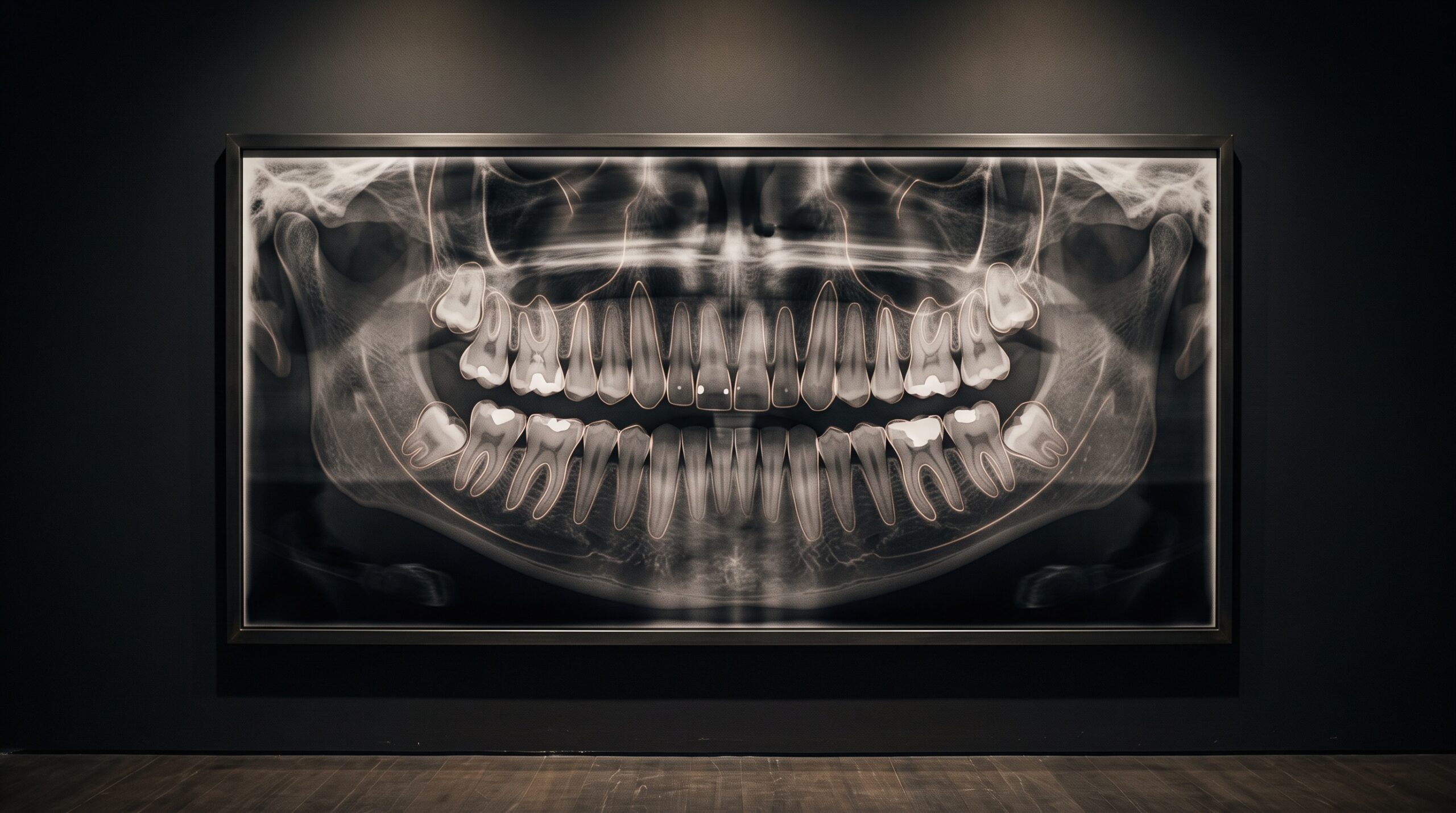
A full dental panoramic radiograph hung as a framed gallery portrait, with faint glowing outlines tracing each tooth as if being read by light

24 Aug One Portrait, Many Verdicts: When a Single Network Reads the Whole Mouth at Once

Posted at 11:38h in Dental Imaging by Raymond 0 Comments
0 Likes

Most dental AI has been trained to answer one question at a time. A newer class of models reads the entire panoramic as a single portrait and returns many verdicts at once - and the craft that makes that possible is quieter than the network...

Read More
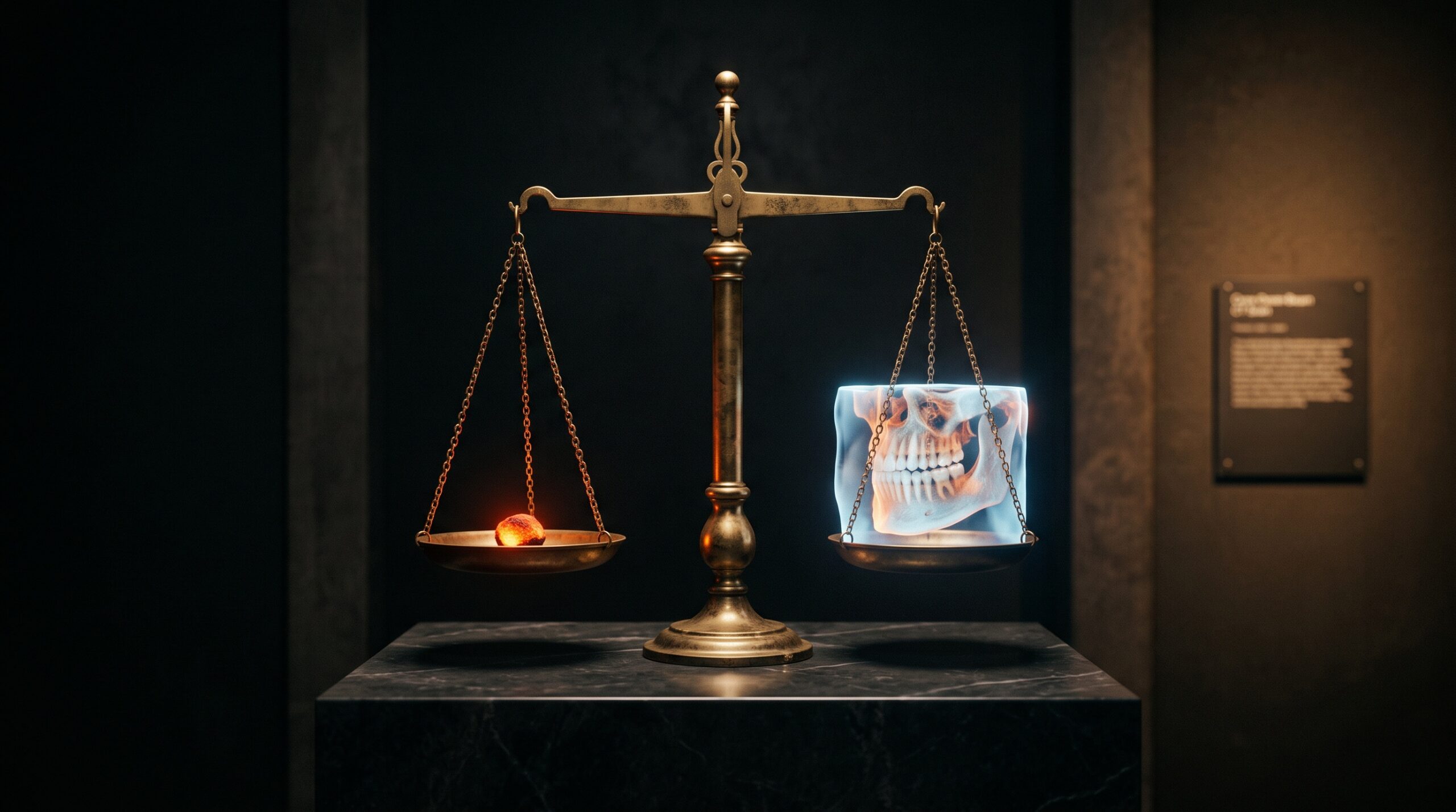
A brass balance scale in a gallery, one pan holding a glowing ember of radiation, the other a luminous 3D jaw scan, hanging near balance

23 Aug The Risk of Not Looking: Weighing a Cone-Beam Scan’s Radiation Against What It Reveals

Posted at 10:39h in Dental Imaging by Raymond 0 Comments
0 Likes

We are trained to fear the dose. We are rarely trained to fear the missed diagnosis. A cone-beam scan is a decision between two risks, not one - and the picture that goes unmade has a cost of its own....

Read More
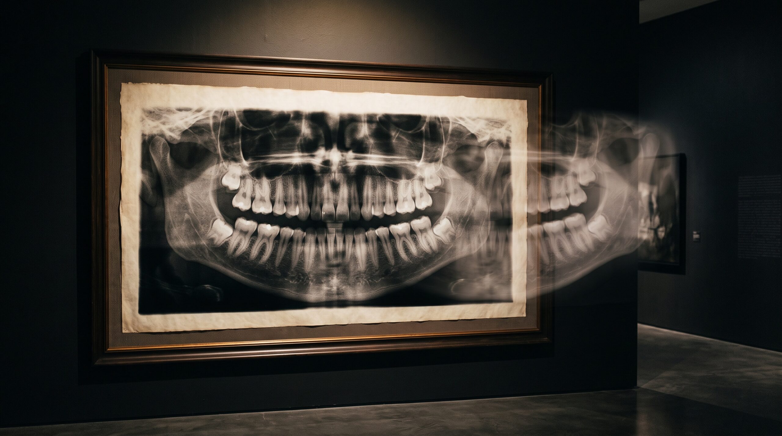
A framed panoramic dental radiograph on a gallery wall with a faint translucent ghost shadow floating over the opposite side of the jaw

22 Aug The Ghost on the Other Side: How a Panoramic X-Ray Paints a Shadow Where No Bone Exists

Posted at 11:31h in Dental Imaging by Raymond 0 Comments
0 Likes

There is a second image hiding inside every panoramic radiograph: faint, doubled, floating on the wrong side of the jaw. It is not a machine fault. It is geometry - and learning to read it is a quiet mark of the craft....

Read More
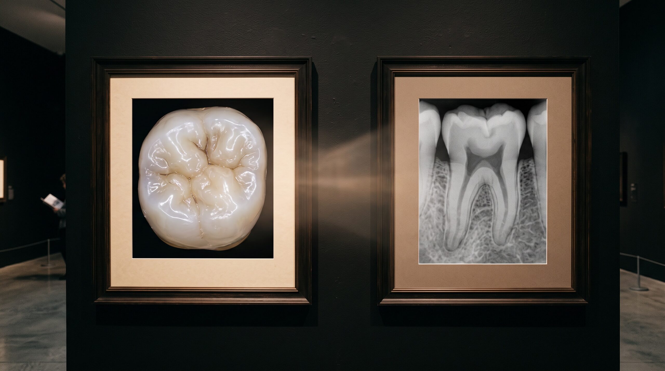
Two framed images of the same molar side by side - a color surface scan and a grey radiograph - with light suggesting they are merging

21 Aug Two Ways of Seeing, One Frame: Fusing the Intraoral Scan and the Radiograph Into a Single Image

Posted at 10:38h in Dental Imaging by Raymond 0 Comments
0 Likes

One instrument captures the surface and refuses to look beneath it. The other looks straight through and forgets the surface exists. For decades the clinician fused them in the mind. Now the software does it - and the craft is changing....

Read More
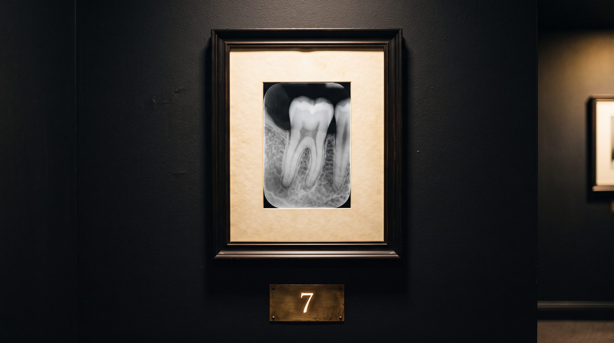
A framed dental radiograph on a gallery wall with a small brass placard beneath it bearing a single glowing number

20 Aug The Dose You Cannot See: Exposure Index, Dose Creep, and the Number a Forgiving Sensor Hides

Posted at 11:39h in Dental Imaging by Raymond 0 Comments
0 Likes

Film punished overexposure by going dark. Digital rewards it with a cleaner picture - which is exactly how dose creeps upward, invisibly. Here is the number that fixes it....

Read More
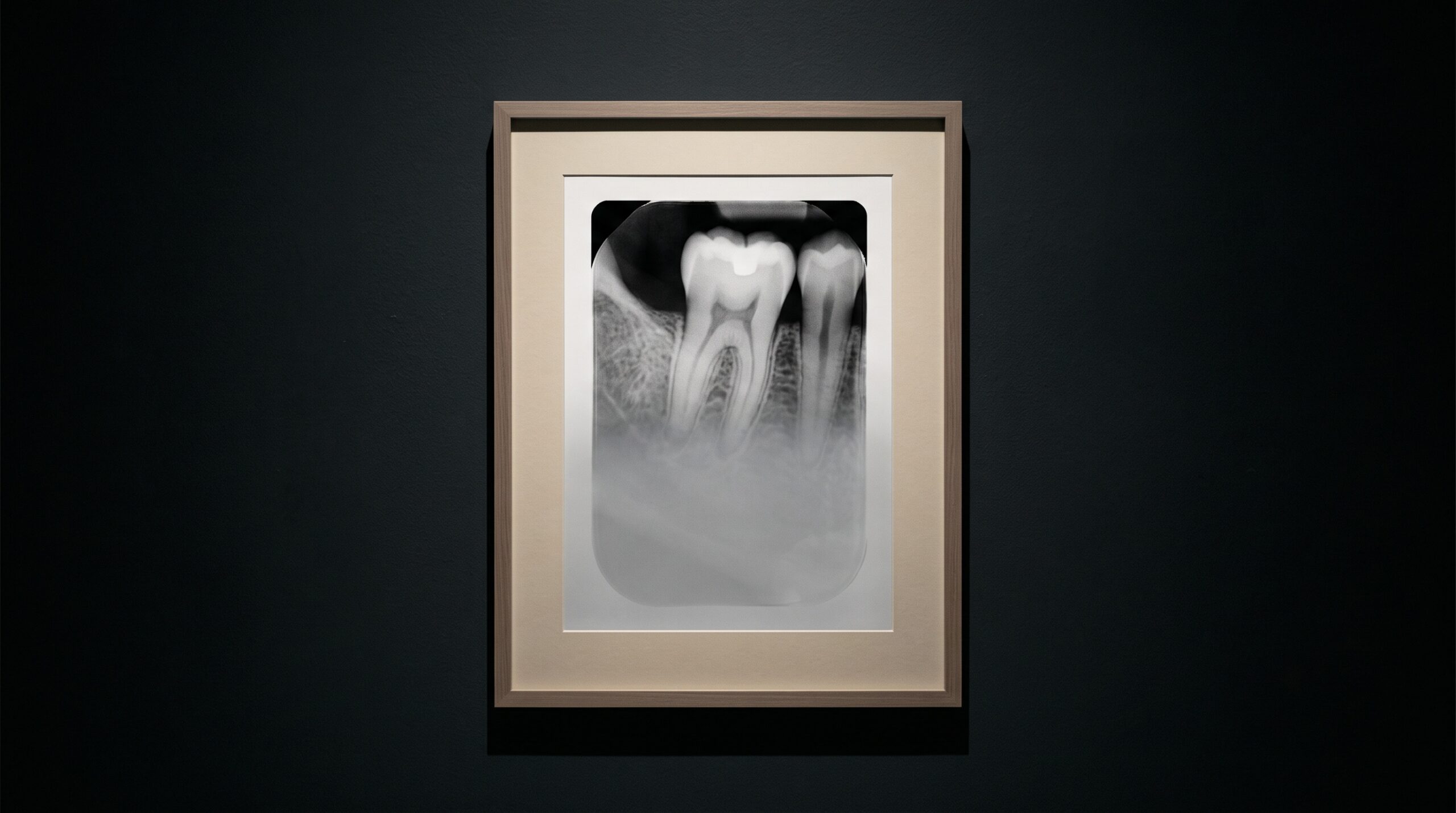
A framed dental radiograph with a soft translucent grey veil drifting over its lower half, washing out the contrast there

19 Aug The Scattered Photon: How Compton Scatter, Collimation, and the Missing Grid Shape Contrast in Intraoral Radiography

Posted at 10:37h in Dental Imaging by Raymond 0 Comments
0 Likes

A radiograph's contrast is built by the photons that are cleanly absorbed and the ones that pass straight through. The scattered photon belongs to neither, and it is the quiet vandal of the image - a fog with no source that dental imaging fights not...

Read More
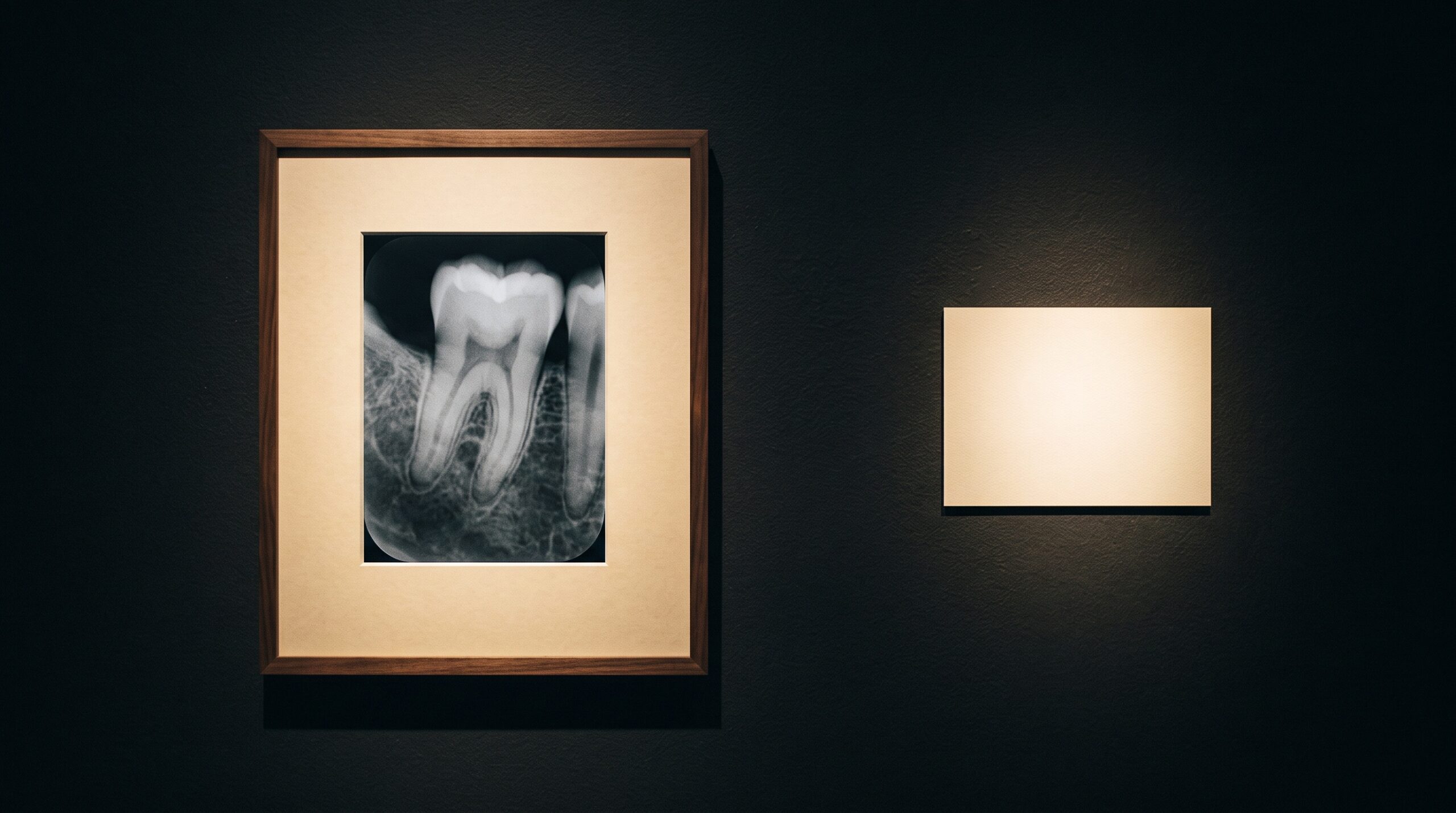
A framed dental radiograph on a gallery wall with a small blank museum placard beside it under refined lighting

18 Aug The Placard Beside the Picture: How Structured Reporting Makes a Dental Radiograph Legible

Posted at 10:38h in Dental Imaging by Raymond 0 Comments
0 Likes

In a gallery, the placard beside the artwork decides how much of the work you actually take in. The radiograph has a placard too - the report - and a new comparative study shows that giving it a fixed structure, rather than free-flowing prose, measurably...

Read More
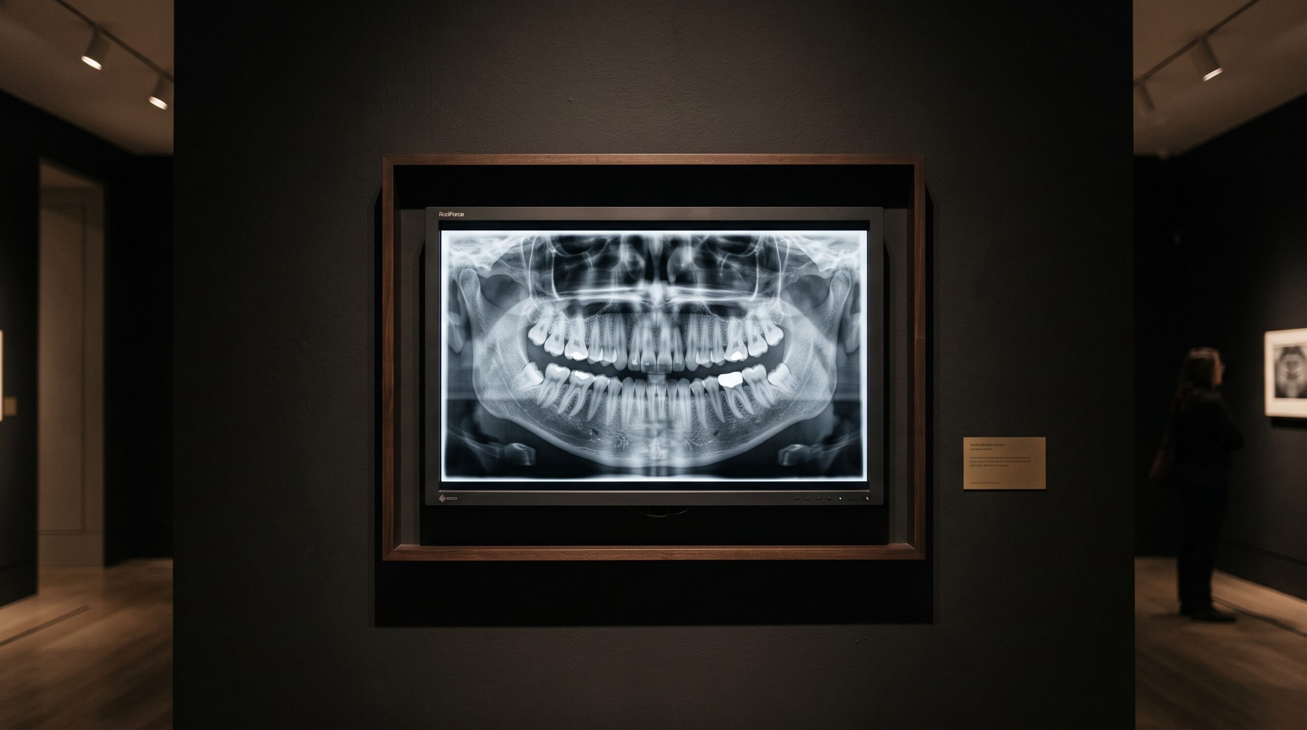
A diagnostic display monitor shown as framed gallery art, its screen holding a dental radiograph in a smooth ramp of grays

17 Aug The Last Instrument: Diagnostic Display Calibration and the DICOM Grayscale Standard for Reading a Radiograph

Posted at 11:38h in Dental Imaging by Raymond 0 Comments
0 Likes

Every radiograph ends its journey on a screen, and the screen is the one instrument in the imaging chain that most practices never calibrate. The DICOM grayscale standard exists because the human eye reads brightness unevenly — and because a monitor that does not account...

Read More
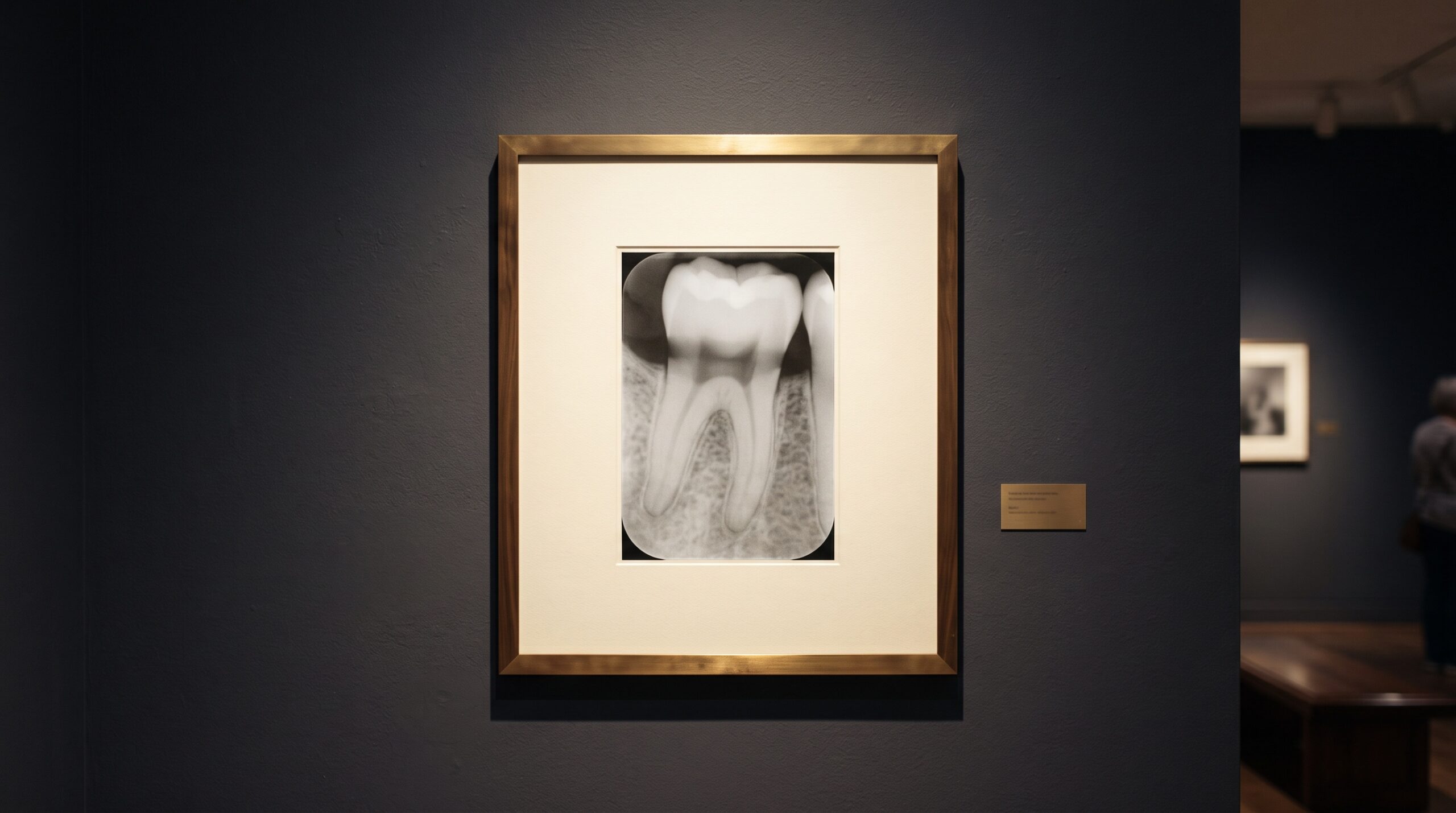
A framed dental radiograph under gallery lighting showing a soft diffuse dark band at the neck of a tooth between crown and bone

16 Aug Cervical Burnout: The Radiolucent Illusion at the Tooth’s Neck That Mimics Caries

Posted at 13:39h in Dental Imaging by Raymond 0 Comments
0 Likes

Between the bright cap of the enamel and the pale crest of the bone runs a soft dark band along the neck of many teeth. It can look, unmistakably, like decay eating into the root. Often it is nothing of the kind — it is...

Read More
  • 1
  • 2
  • 3
  • 4
  • 5
  • 6
  • 7
  • 8
  • 9
  • 10
  • 11
  • 12
  • 13
  • 14
  • 15
© Copyright 2020, Raster Builders Inc. All Rights Reserved.