Logo Logo Logo Logo Logo
  • Home
  • Download
  • About
  • Blog
  • Home
  • Download
  • About
  • Blog
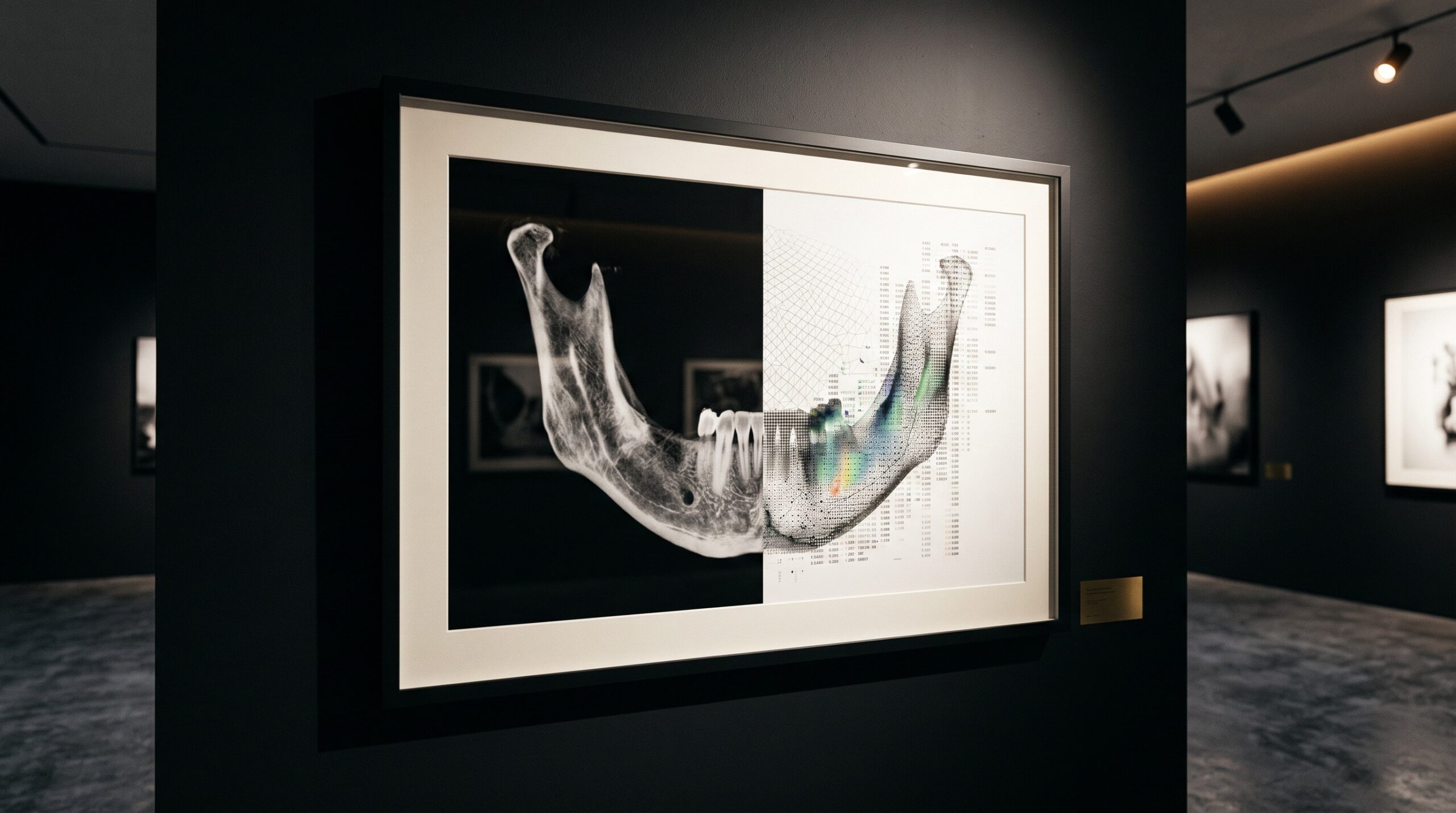
A framed CBCT cross-section of the jaw, one half a bone image and the other half dissolving into a faint numeric grid, presented as gallery art

09 Oct The Numbers Beneath the Picture: Radiomics and the Quantitative Dental Image

Posted at 19:17h in Dental Imaging by Raymond 0 Comments
0 Likes

A radiograph is also a field of numbers. Dental radiomics mines the texture a trained eye cannot name — trabecular patterns, gray-level statistics, subtle lesions — and turns the image into a measurable biomarker. A look at what radiomic features are, what recent dental studies...
Read More
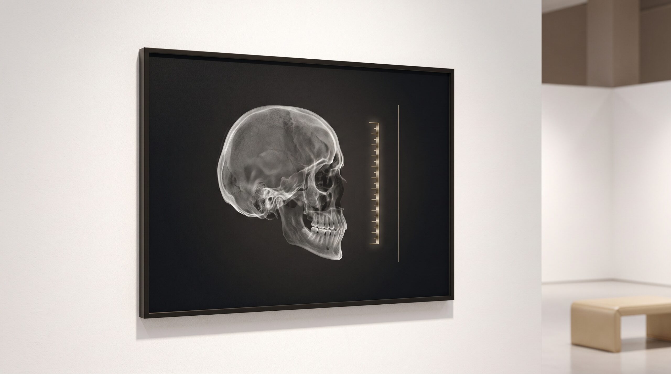
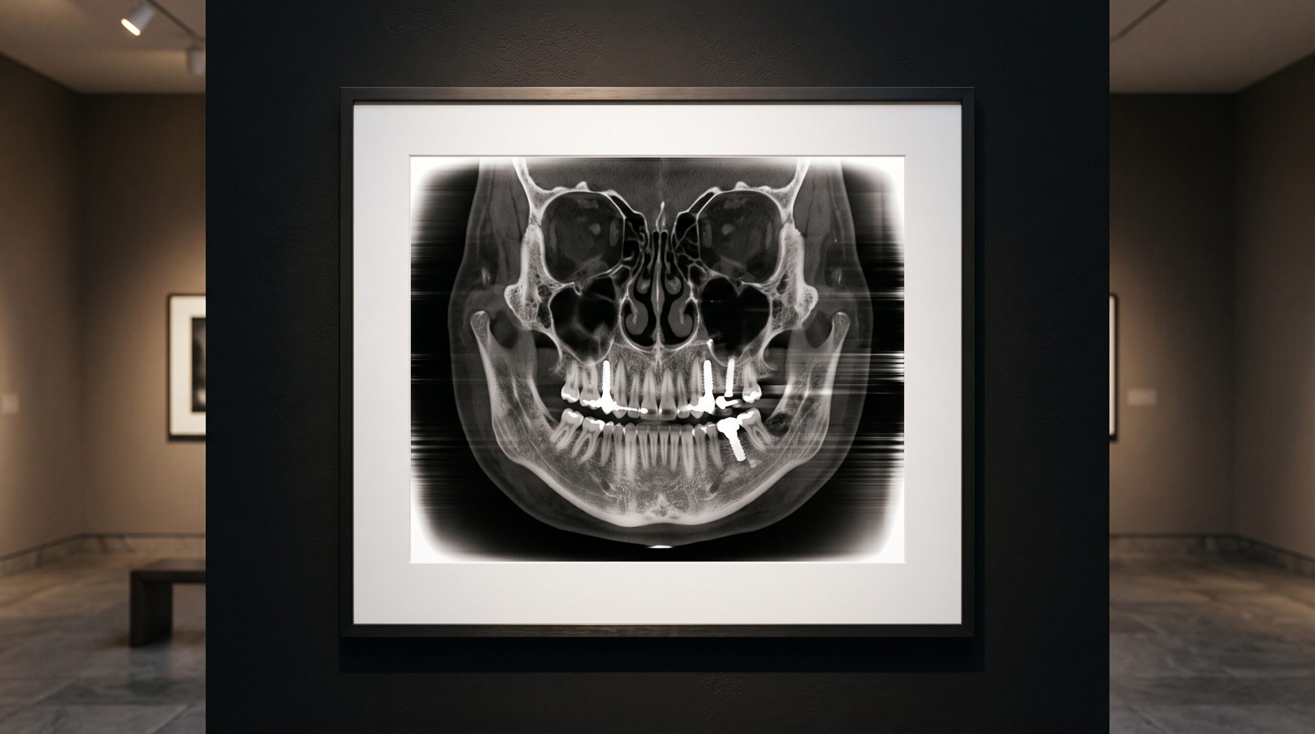
A framed cone-beam CT cross-section of the skull with bright cupping and streaks at its edges, presented as gallery art

08 Oct Painting Back the Edge of the Scan: How Diffusion AI Erases CBCT Truncation Artifacts

Posted at 12:22h in Dental Imaging by Raymond 0 Comments
0 Likes

When a patient's anatomy spills past the edge of a cone-beam CT detector, the reconstruction cups and streaks where the data ran out. A new conditional diffusion model restores those missing measurements in the raw projections, delivering markedly cleaner scans with no new hardware and...

Read More
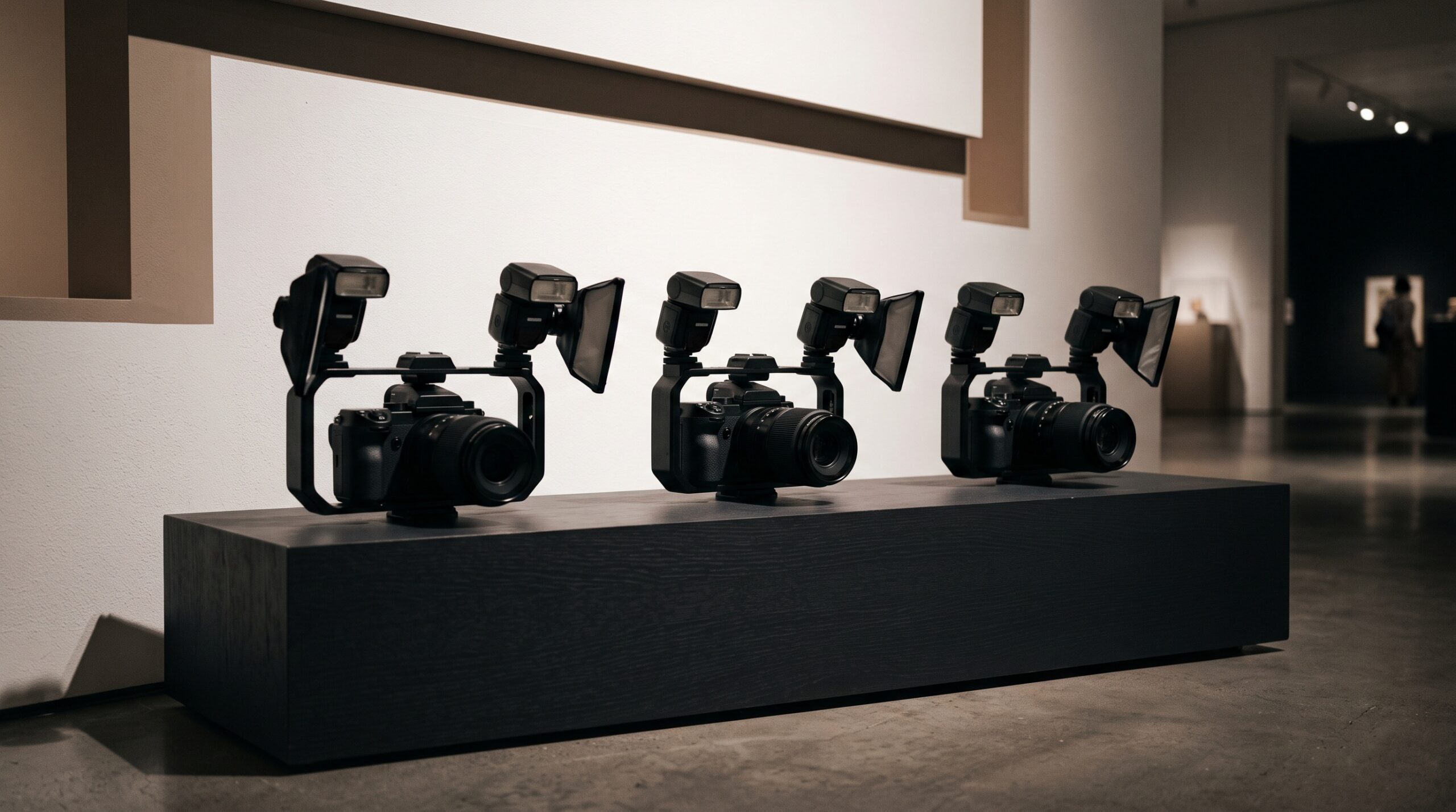
Three mirrorless cameras fitted with macro lenses and twin-flash brackets displayed as gallery sculptures

07 Oct The Best Mirrorless Cameras for Dental Photography in 2026

Posted at 11:31h in Clinical Photography by Raymond 0 Comments
0 Likes

The best camera for dental photography is rarely decided by the body. It is decided by the macro lens, the flash, and the hand behind them. Here is an honest, criteria-scored roundup of the mirrorless systems worth building a dental kit around in 2026 —...

Read More
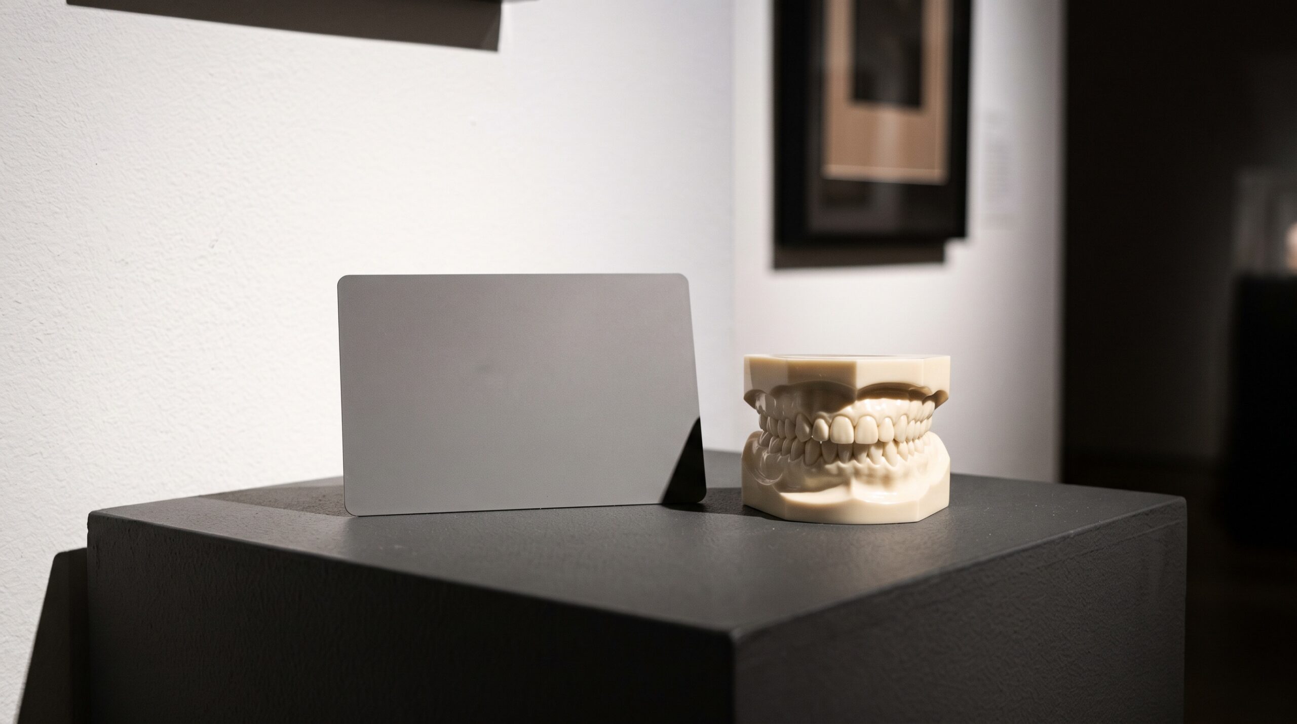
A neutral gray reference card beside a dental study model under refined gallery lighting

06 Oct The Gray Card in the Operatory: Nailing White Balance at the Moment of Capture

Posted at 12:41h in Clinical Photography by Raymond 0 Comments
0 Likes

Color accuracy in dental photography is won or lost in the fraction of a second the shutter opens. A neutral gray card, placed in the frame and used to set a custom white balance, is the quiet discipline that lets a ceramist trust the color...

Read More
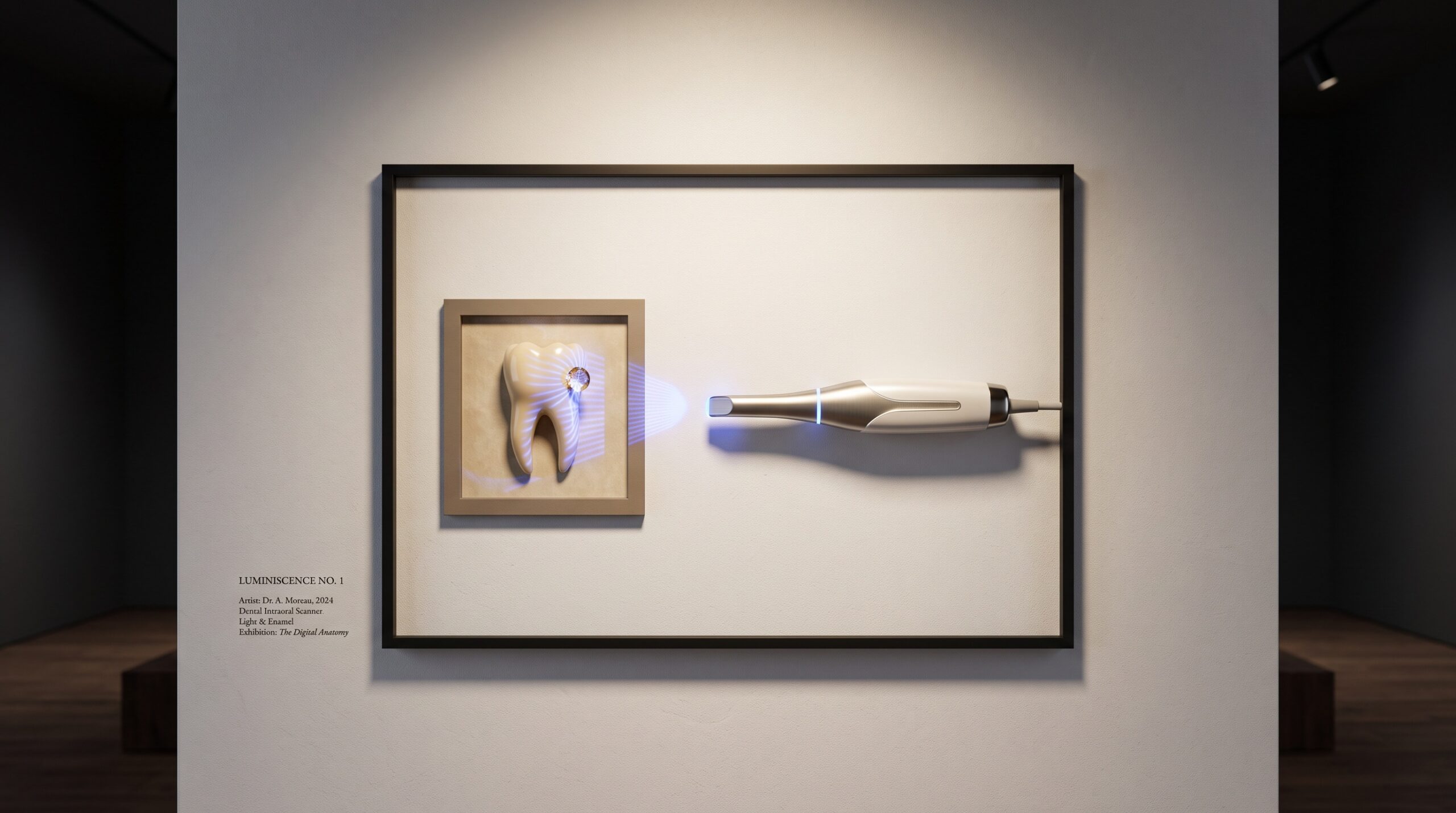
An intraoral scanner tip casting structured light on a framed tooth with a single bright bead of liquid on the enamel

05 Oct Keeping It Dry: How Moisture Control Governs Intraoral Scan Accuracy

Posted at 17:03h in Dental Imaging by Raymond 0 Comments
0 Likes

An intraoral scanner is an optical instrument pointed at the wettest surface in medicine. A film of saliva, a bead of blood at the margin, a breath of fog on the tip — each is invisible on the monitor and quietly fatal to the fit....

Read More
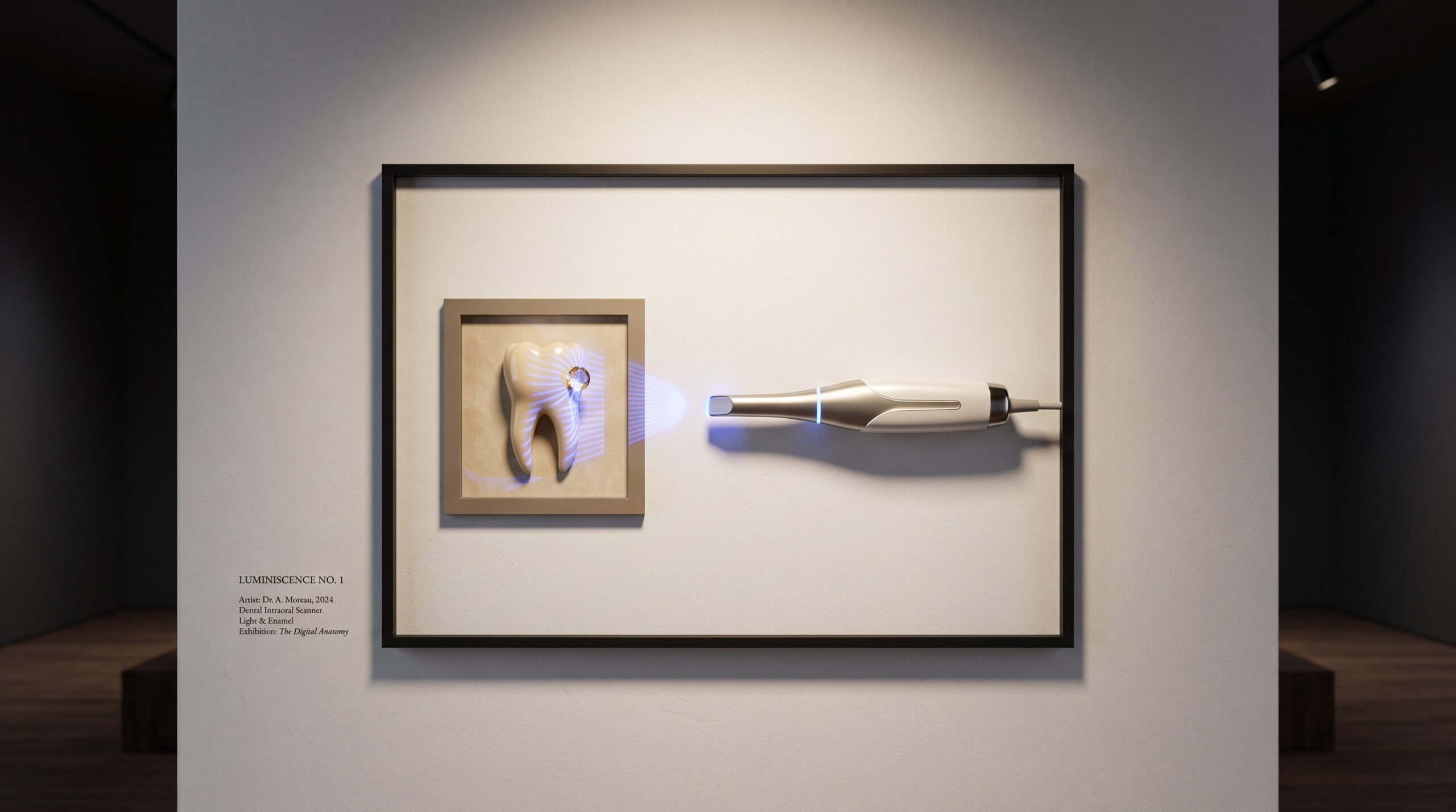
An intraoral scanner tip casting structured light on a framed tooth with a single bright bead of liquid on the enamel

05 Oct Keeping It Dry: How Moisture Control Governs Intraoral Scan Accuracy

Posted at 17:02h in Dental Imaging by Raymond 0 Comments
0 Likes

An intraoral scanner is an optical instrument pointed at the wettest surface in medicine. A film of saliva, a bead of blood at the margin, a breath of fog on the tip — each is invisible on the monitor and quietly fatal to the fit....

Read More
A grey CBCT skull volume framed as gallery art beside a glowing measured threshold line

04 Oct How Much Is Normal? Diagnostic Reference Levels and the Quiet Discipline of CBCT Dose

Posted at 10:36h in Dental Imaging by Raymond 0 Comments
0 Likes

A cone-beam scanner will give you the dose you ask of it, and it never asks whether you needed that much. A Diagnostic Reference Level is the first honest answer to a question most practices never pose: compared with everyone else doing this exact exam,...

Read More
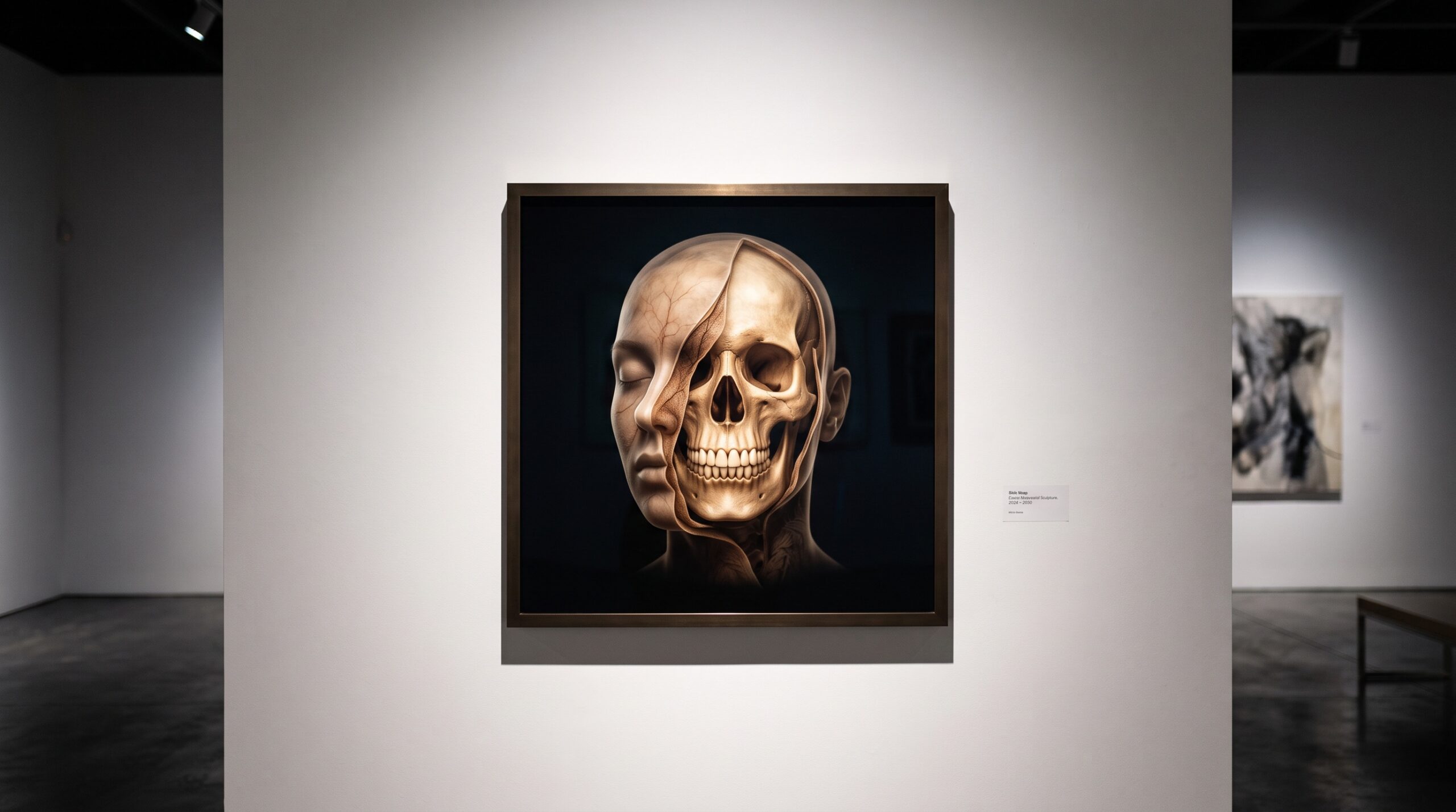
Composite 3D model of a head showing translucent face over aligned skull and teeth, framed as gallery art

03 Oct The Virtual Patient: Fusing Face Scans, CBCT, and Intraoral Data Into One 3D Record

Posted at 10:40h in Dental Imaging by Raymond 0 Comments
0 Likes

A face scan knows the smile but not the bone. A CBCT knows the bone but not the skin. An intraoral scan knows the teeth to the micron but nothing above the gumline. Fuse all three into one aligned 3D record and you have something...

Read More
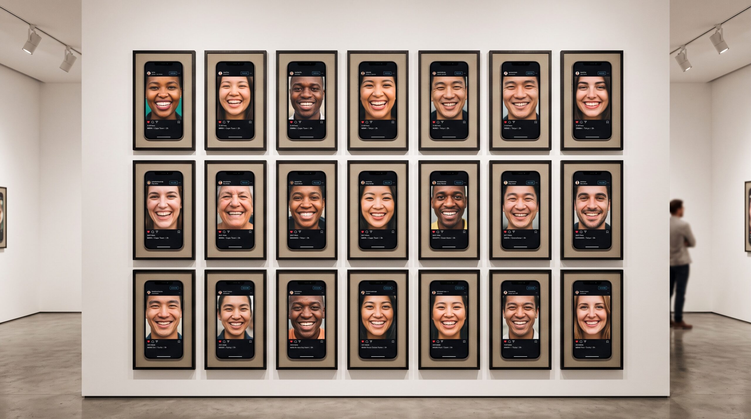
A gallery wall of framed smartphone-shaped smile photographs arranged like a curated social feed

02 Oct The Feed as Gallery: The Aesthetics, Ethics, and Consent of Dental Photography on Social Media

Posted at 12:21h in Clinical Photography by Raymond 0 Comments
0 Likes

A clinical photograph made for the chart and a clinical photograph made for the feed are no longer the same object. When a patient's smile becomes public content, the quiet craft of documentation acquires an audience — and a conscience....

Read More
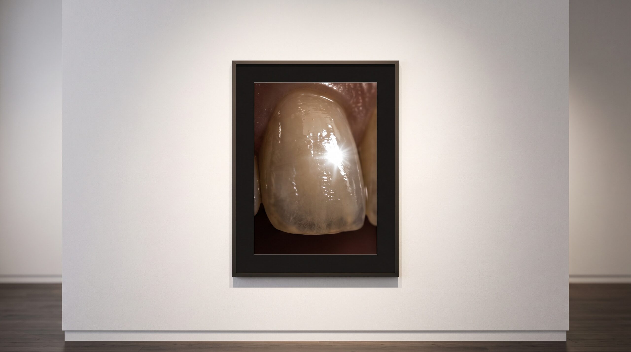
Macro photograph of a tooth with a bright specular highlight on wet enamel, framed as gallery art

01 Oct The Highlight That Burns White: HDR, Exposure Bracketing, and the Craft of Photographing Wet Enamel

Posted at 10:39h in Clinical Photography by Raymond 0 Comments
0 Likes

Polished, saliva-wet enamel throws a specular highlight so bright it burns to pure white, while the back of the arch falls into shadow. Exposure bracketing and HDR promise to hold both at once — if you can keep the result honest....

Read More
  • 1
  • 2
  • 3
  • 4
  • 5
  • 6
  • 7
  • 8
  • 9
  • 10
  • 11
  • 12
  • 13
  • 14
  • 15
  • 16
  • 17
  • 18
  • 19
  • 20
© Copyright 2020, Raster Builders Inc. All Rights Reserved.