Logo Logo Logo Logo Logo
  • Home
  • Download
  • About
  • Blog
  • Home
  • Download
  • About
  • Blog
A luminous colour 3D digital model of a child's dental arch displayed as gallery art, one tooth showing a faint chalky white lesion

11 Sep The Mouth on the Screen: Diagnosing Childhood Decay From a 3D Intraoral Scan

Posted at 10:39h in Dental Imaging by Raymond 0 Comments
0 Likes

For a century, diagnosing decay in a small child's mouth has meant one thing: a clinician, a mirror, a light, and a cooperative patient sitting still long enough to be examined. A 2025 Australian study quietly proposed an alternative. Capture a colour three-dimensional scan of...

Read More
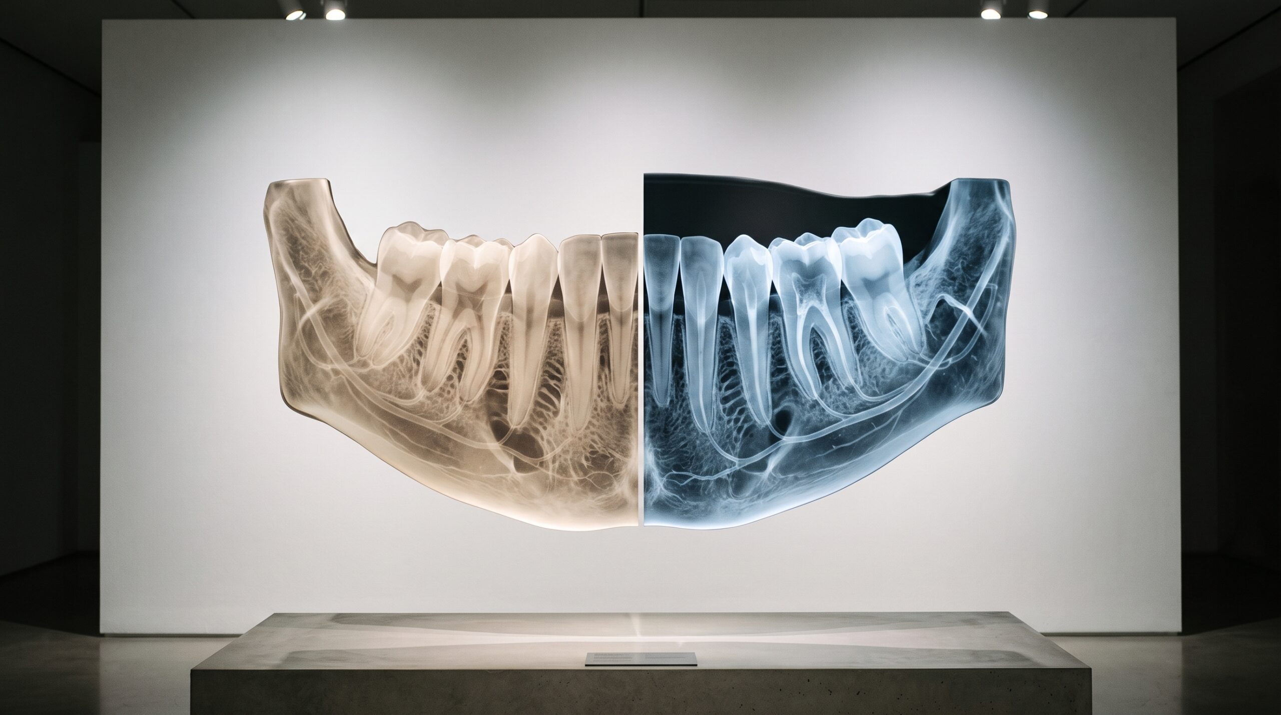
A gallery triptych: two near-identical bone radiographs and a third flat-grey panel showing only the glowing region where they differ

10 Sep The Art of Seeing Change: Digital Subtraction Radiography and Imaging What the Eye Cannot

Posted at 11:30h in Dental Imaging by Raymond 0 Comments
0 Likes

Two radiographs of the same tooth, taken months apart, can look identical to a trained eye while a real lesion quietly advances between them. Digital subtraction radiography is the technique that makes that invisible change visible: align the two images perfectly, subtract one from the...

Read More
A single empty ornate frame on a gallery wall, evoking the radiograph deliberately not taken

09 Sep The Discipline of Restraint: The New Imaging Guidance and the Art of the Radiograph Not Taken

Posted at 10:39h in Dental Imaging by Raymond 0 Comments
0 Likes

The most revealing decision a clinician makes about a radiograph is often whether to take it at all. The first ADA and AAOMR patient-selection recommendations in more than a decade - and the first to address cone-beam CT - reframe imaging not as a schedule...

Read More
A framed lateral skull X-ray displayed as gallery art, with small glowing points marking anatomical landmarks along the bone profile

08 Sep Landmarks by Machine: How AI Traces a Cephalometric X-Ray for Orthodontic Analysis

Posted at 10:37h in Dental Imaging by Raymond 0 Comments
0 Likes

A lateral cephalogram is one of the most standardized images in all of dentistry - a skull rendered in exact geometry so that the same points can be found on it year after year. For a century, finding those points was patient handwork with a...

Read More
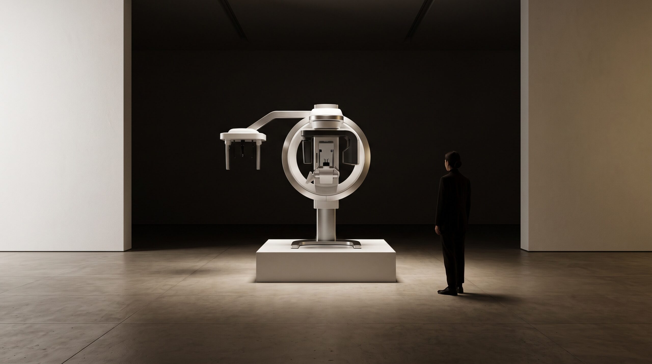
A handheld dental X-ray unit with a round backscatter shield at the cone, held as if it were a piece of gallery sculpture

07 Sep Can a Handheld Radiograph Match the Wall-Mount? The Physics of Image Quality in Portable Dental X-Ray

Posted at 10:38h in Dental Imaging by Raymond 0 Comments
0 Likes

A wall-mounted X-ray head is a fixed instrument, aligned by a stable arm and switched from behind a wall. A handheld unit puts the same tube in one gloved hand and asks the clinician to be both aimer and tripod. The device has become common...

Read More
A dental photogrammetry marker abutment displayed as sculpture, fine lines of light triangulating onto it from several directions

06 Sep Triangulating the Arch: How Photogrammetry Solves the Full-Mouth Implant Impression

Posted at 10:39h in Dental Imaging by Raymond 0 Comments
0 Likes

For a single crown, an intraoral scanner is enough. For a full arch of implants, the errors of scanning add up across the span until the bridge no longer fits the bone it was built for. Photogrammetry answers with a different discipline entirely: it does...

Read More
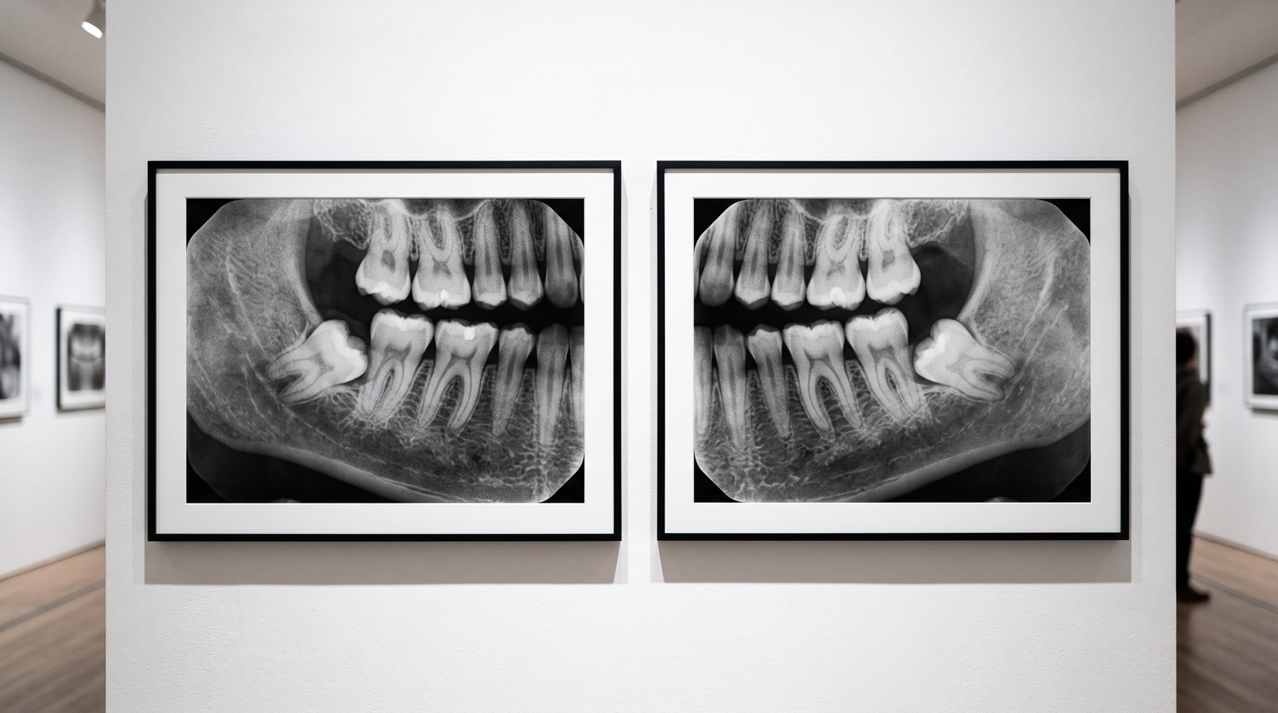
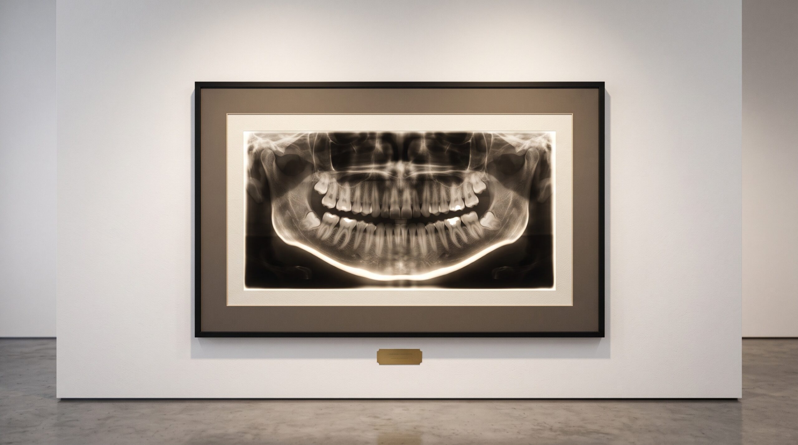
A panoramic dental radiograph framed as fine art, the frame deliberately wider than the teeth to include the surrounding anatomy

05 Sep The Whole Picture: What a Dental X-Ray Reveals Beyond the Teeth

Posted at 10:38h in Dental Imaging by Raymond 0 Comments
0 Likes

A dental radiograph is framed for your teeth, but the exposure does not stop at the enamel. In the same grey field sit the sinuses, the jaw, the airway, the soft tissues of the neck - and now and then, something no one was looking...

Read More
Three dental x-ray detectors displayed as artworks on brass museum plinths: two rigid corded solid-state sensors and one thin flexible phosphor plate.

04 Sep Three Ways to Catch a Photon: CCD, CMOS, and Phosphor Plates Compared

Posted at 10:39h in Dental Imaging by Raymond 0 Comments
0 Likes

Every digital intraoral image begins with a decision made in silicon or phosphor: how to turn an x-ray photon into a number. There are three answers in common use, and each one quietly shapes what the radiograph can and cannot show....

Read More
Two framed versions of the same dental radiograph on a gallery wall, one crisp and one muddy and underexposed

04 Sep Garbage In, Garbage Out: Image Quality and Why AI Still Needs a Good Radiograph

Posted at 09:44h in Dental Imaging by Raymond 0 Comments
0 Likes

A detection model will read an underexposed, cone-cut, motion-blurred bitewing without hesitation and hand you a confident, tidy result. That composure is the danger. Everything AI concludes about a radiograph is downstream of how well the image was made - and the machine cannot see...

Read More
A single pale cube on a brass museum plinth engraved with a number that appears to shimmer and double, presented as an artwork whose value cannot be trusted.

03 Sep The Number That Lies: Why a CBCT’s Gray Values Are Not True Hounsfield Units

Posted at 10:37h in Dental Imaging by Raymond 0 Comments
0 Likes

On a medical CT, the number in a voxel is a promise: water reads zero, cortical bone reads a thousand, and the scale means the same thing tomorrow as today. On a cone-beam scan, the same-looking number keeps no such promise - and mistaking one...

Read More
  • 1
  • 2
  • 3
  • 4
  • 5
  • 6
  • 7
  • 8
  • 9
  • 10
  • 11
  • 12
© Copyright 2020, Raster Builders Inc. All Rights Reserved.