Logo Logo Logo Logo Logo
  • Home
  • Download
  • About
  • Blog
  • Home
  • Download
  • About
  • Blog

11 Feb Understanding AI in Dental Imaging: How Artificial Intelligence Is Changing Radiograph Analysis

Posted at 14:09h in Dental Imaging by Raymond 0 Comments
1 Like

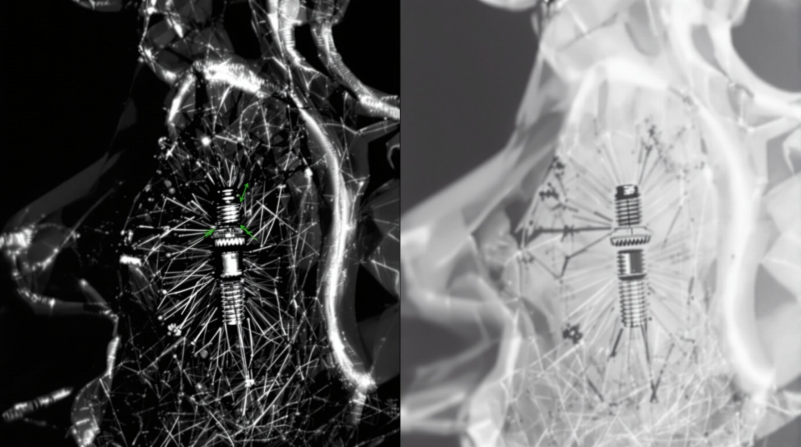
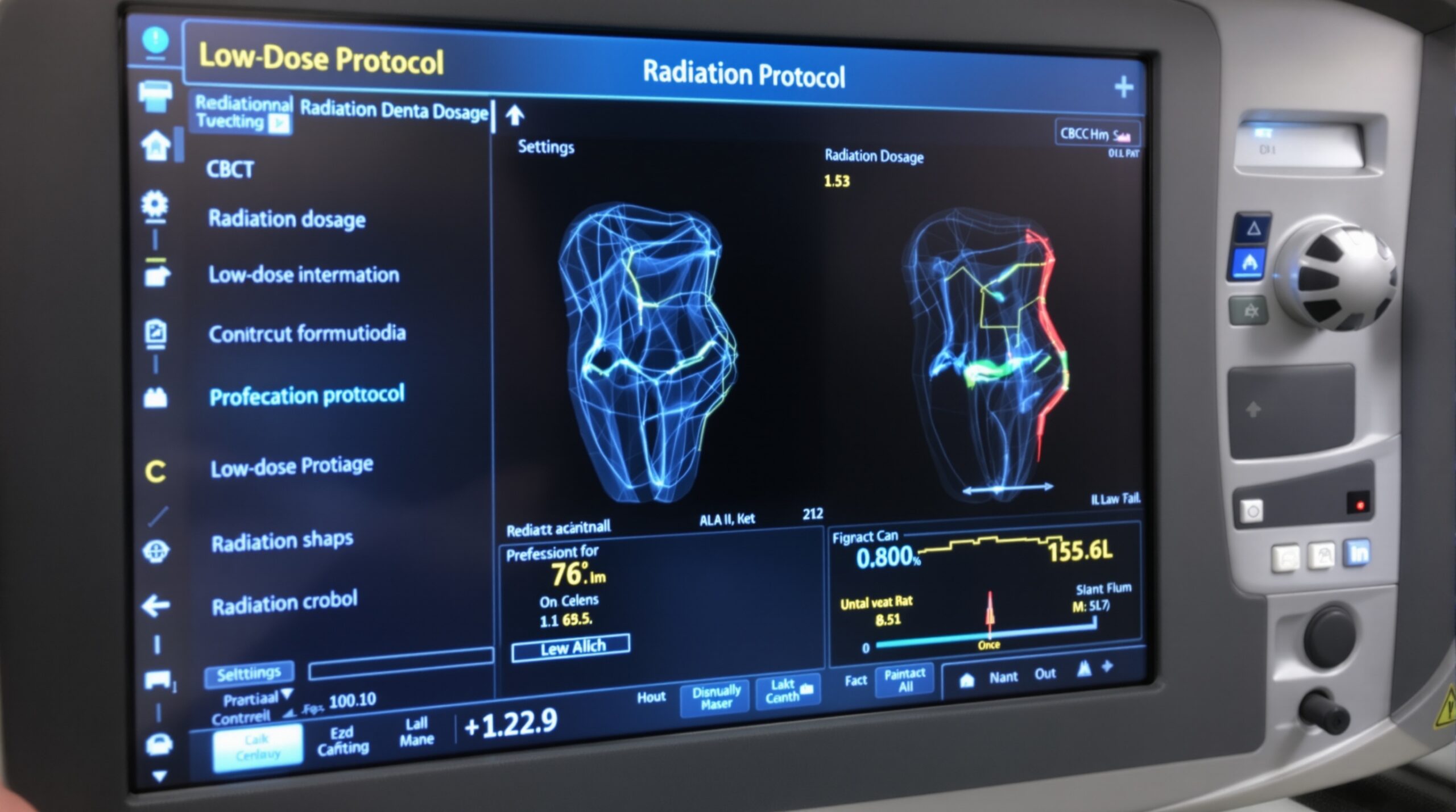
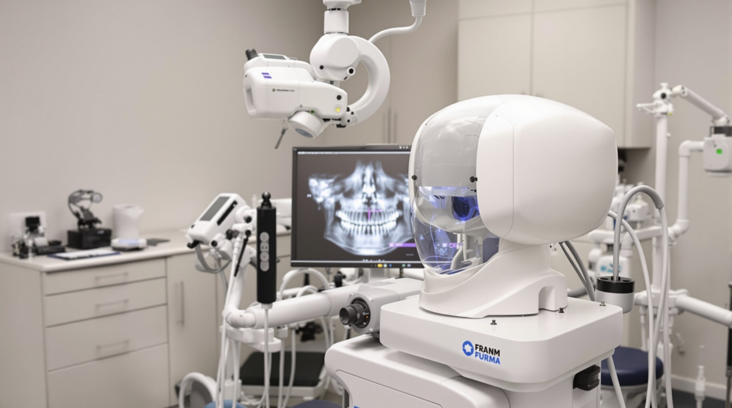
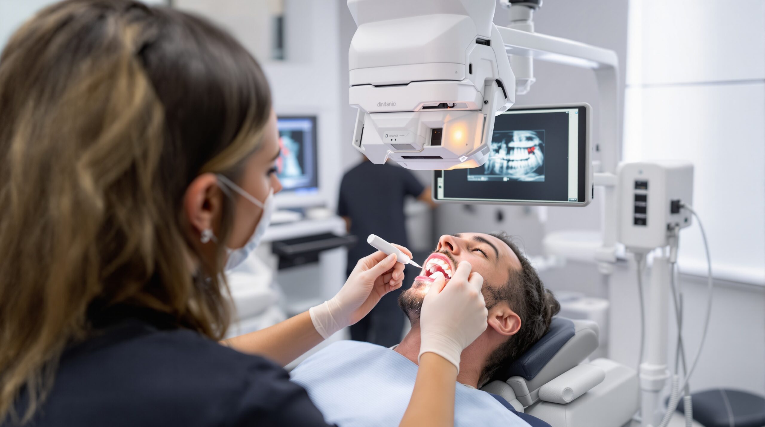
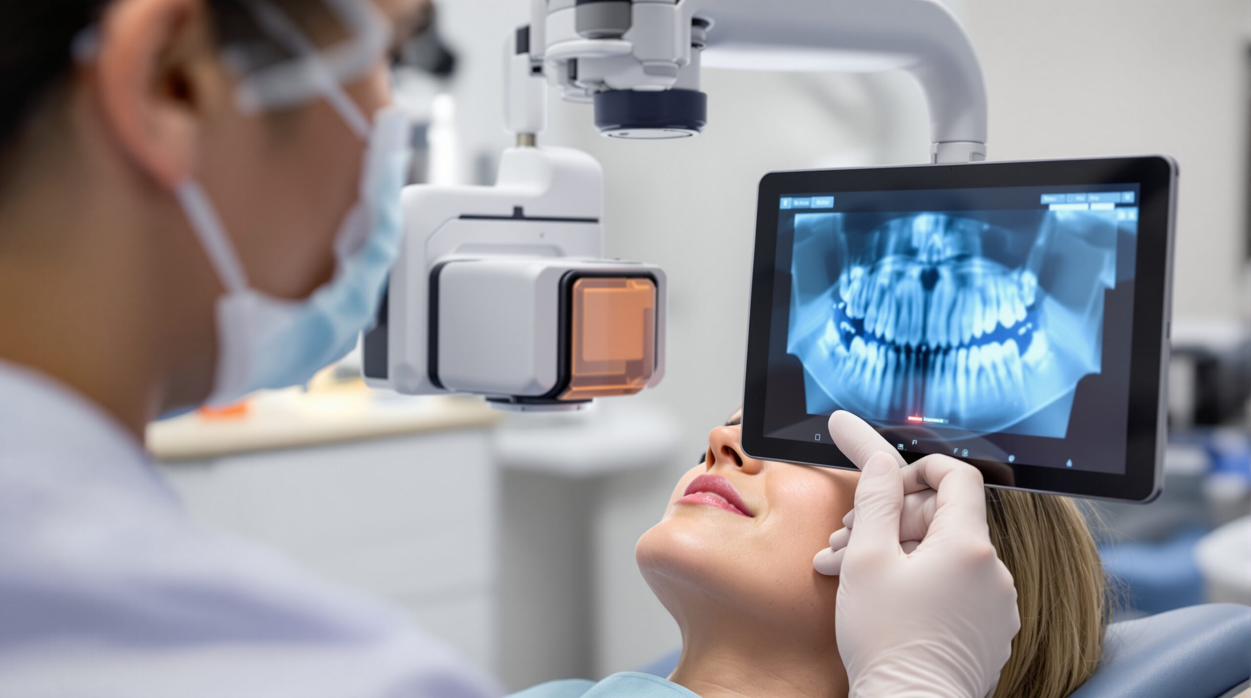
Artificial intelligence is rapidly transforming dental imaging. From detecting early-stage cavities that the human eye might miss to analyzing complex 3D CBCT scans, AI-powered tools are becoming a practical reality in dental practices around the world. But what exactly can AI do today, where are...

Read More
A framed dental periapical radiograph on a gallery wall beside a glowing panel appraising it with a faint scoring overlay

09 Feb When the Machine Becomes the Critic: Deep Learning Now Grades the Radiograph Itself

Posted at 10:12h in Dental Imaging by Raymond 0 Comments
0 Likes

A neural network can now look at a periapical radiograph and tell you, in the instant it is captured, whether it is diagnostic. What it means when the image acquires its own critic....

Read More
A dental radiograph presented as framed fine art on a gallery wall, its background deep true-black

08 Feb The Photon-Counting Detector: A New Physics Beneath the Radiograph

Posted at 10:12h in Dental Imaging by Raymond 0 Comments
0 Likes

For a hundred years the dental radiograph has been an average — a blur of charge summed across an exposure. The photon-counting detector refuses to average. It counts each X-ray individually, and in doing so quietly rewrites what a diagnostic image can be....

Read More
A dimensionally faithful dental periapical radiograph presented as framed fine art on a gallery wall

07 Feb The Geometry of an Honest Radiograph: Paralleling, the Bisecting Angle, and the Truth About Length

Posted at 10:12h in Dental Imaging by Raymond 0 Comments
0 Likes

A dental radiograph looks like a photograph, but it is really a measurement — a shadow cast by an X-ray beam onto a flat receptor. Whether that shadow reports a root's true length or a convincing lie depends almost entirely on geometry, and on the...

Read More
A dental macro photograph with one tooth in sharp focus and neighbours softly blurred, framed as gallery art

06 Feb The Plane of Sharpness: Depth of Field, Diffraction, and Focus Stacking in Clinical Dental Photography

Posted at 10:12h in Dental Imaging by Raymond 0 Comments
0 Likes

At the magnifications clinical photography demands, sharpness is not a plane but a sliver — often shallower than a single tooth is deep. Everything that makes a dental image trustworthy begins with a decision about the aperture, and everything the aperture cannot solve, focus stacking...

Read More
A digital 3D mesh of a dental arch presented as framed sculpture on a gallery wall

05 Feb Trueness and Precision: What Really Governs the Accuracy of an Intraoral Scan

Posted at 10:12h in Dental Imaging by Raymond 0 Comments
0 Likes

On the monitor a digital impression is seductive: clean, colour, rotatable, apparently perfect. But a scan that looks perfect and a scan that is accurate are not the same claim, and the gap between them is where restorations quietly fail....

Read More
A handheld intraoral X-ray unit with a round backscatter shield, cradled in gloved hands under gallery lighting

04 Feb The Weight in the Hand: Handheld and Wall-Mounted X-Ray, and What the Dose Really Costs

Posted at 10:12h in Dental Imaging by Raymond 0 Comments
0 Likes

A handheld X-ray unit is a beautiful piece of engineering, and it puts the source of the beam in the operator's own hand. The image it makes is identical to the wall-mounted tube's. The dose it delivers to the person holding it is not....

Read More
  • 1
  • 2
  • 3
  • 4
  • 5
  • 6
  • 7
  • 8
© Copyright 2020, Raster Builders Inc. All Rights Reserved.