Logo Logo Logo Logo Logo
  • Home
  • Download
  • About
  • Blog
  • Home
  • Download
  • About
  • Blog
Three dental x-ray detectors displayed as artworks on brass museum plinths: two rigid corded solid-state sensors and one thin flexible phosphor plate.

04 Sep Three Ways to Catch a Photon: CCD, CMOS, and Phosphor Plates Compared

Posted at 10:39h in Dental Imaging by Raymond 0 Comments
0 Likes

Every digital intraoral image begins with a decision made in silicon or phosphor: how to turn an x-ray photon into a number. There are three answers in common use, and each one quietly shapes what the radiograph can and cannot show....
Read More
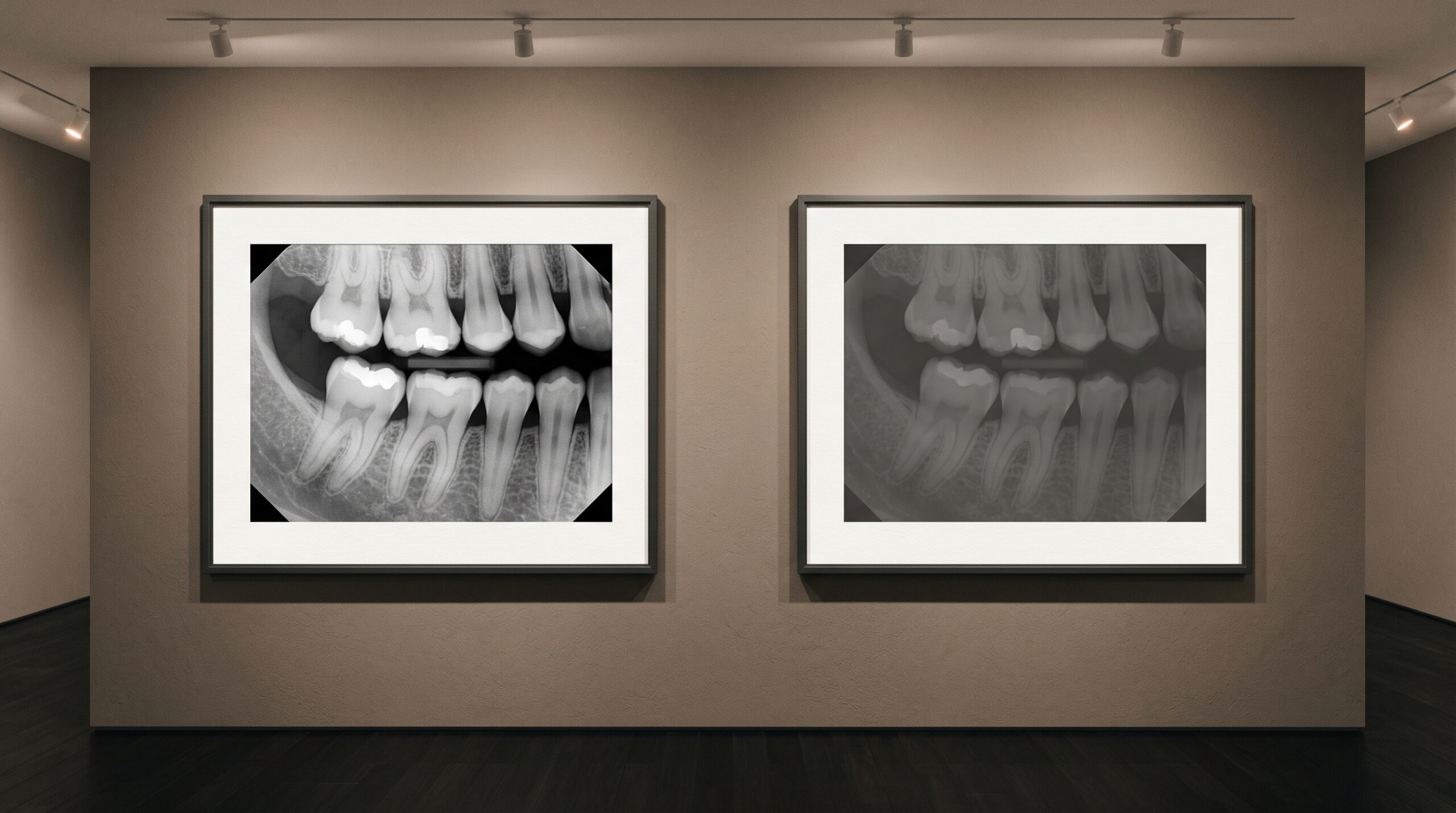
Two framed versions of the same dental radiograph on a gallery wall, one crisp and one muddy and underexposed

04 Sep Garbage In, Garbage Out: Image Quality and Why AI Still Needs a Good Radiograph

Posted at 09:44h in Dental Imaging by Raymond 0 Comments
0 Likes

A detection model will read an underexposed, cone-cut, motion-blurred bitewing without hesitation and hand you a confident, tidy result. That composure is the danger. Everything AI concludes about a radiograph is downstream of how well the image was made - and the machine cannot see...

Read More
A single pale cube on a brass museum plinth engraved with a number that appears to shimmer and double, presented as an artwork whose value cannot be trusted.

03 Sep The Number That Lies: Why a CBCT’s Gray Values Are Not True Hounsfield Units

Posted at 10:37h in Dental Imaging by Raymond 0 Comments
0 Likes

On a medical CT, the number in a voxel is a promise: water reads zero, cortical bone reads a thousand, and the scale means the same thing tomorrow as today. On a cone-beam scan, the same-looking number keeps no such promise - and mistaking one...

Read More
A framed dental radiograph on a gallery wall with soft luminous rings marking a few regions

03 Sep From Detection to Diagnosis: AI-Assisted Radiograph Reporting

Posted at 09:44h in Dental Imaging by Raymond 0 Comments
0 Likes

An AI can mark a dark spot on a bitewing in half a second. Calling that spot a lesion, deciding whether to treat it, and putting your name to the decision are three separate acts the software is not licensed to perform. The report is...

Read More
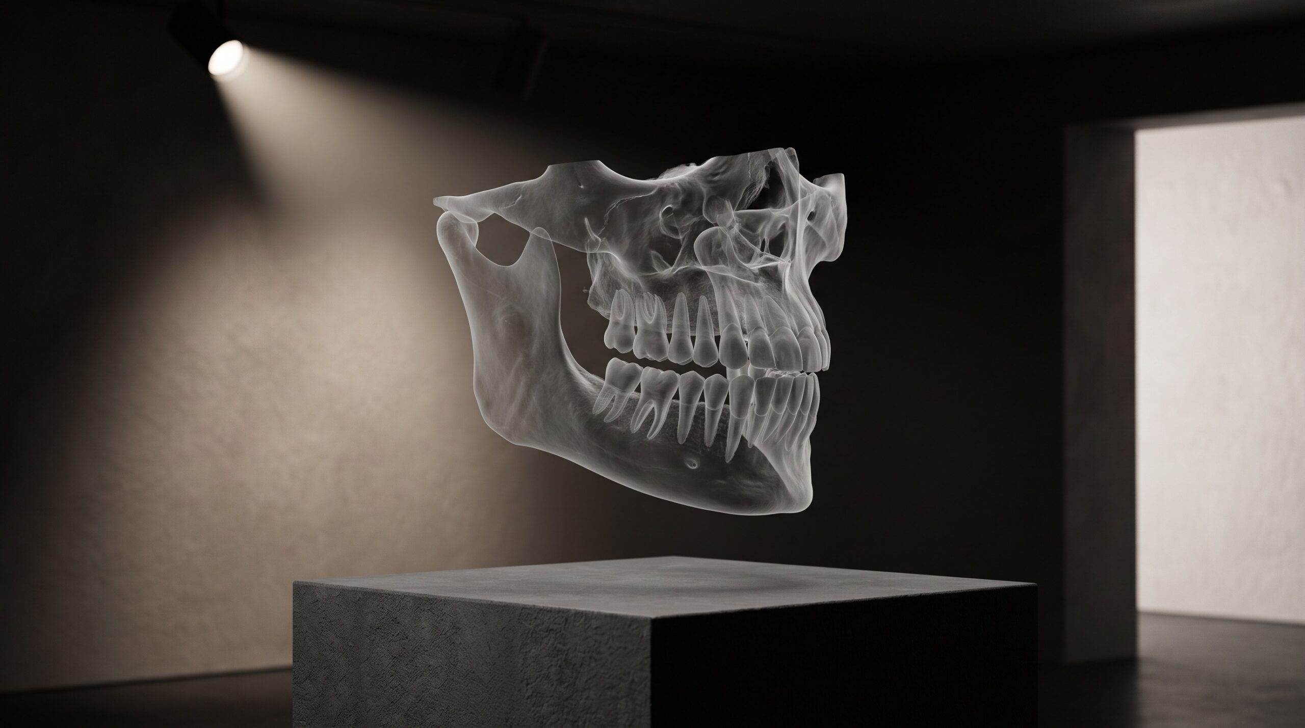
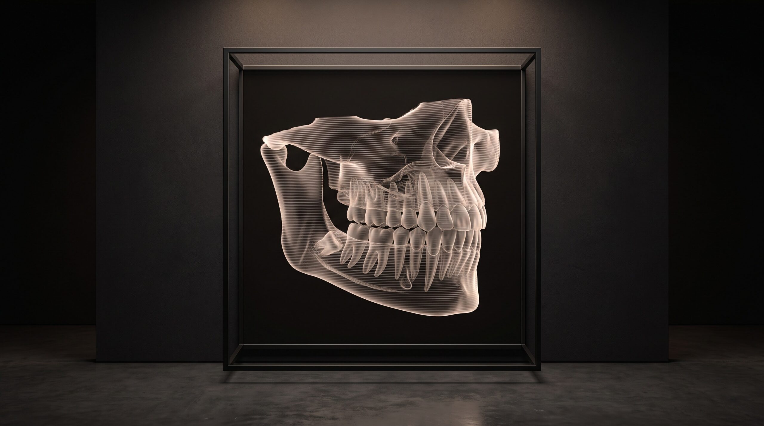
A translucent 3D CBCT volume of a jaw presented as a sculptural exhibit on a gallery plinth

02 Sep AI on CBCT: What 3D Segmentation Actually Finds

Posted at 09:44h in Dental Imaging by Raymond 0 Comments
0 Likes

A CBCT scan arrives as a fog of grey voxels. Segmentation is the act of naming them - drawing the boundary between a tooth and the bone that holds it, between the nerve canal and the space around it. Here is what the machine actually...

Read More
A blank digital x-ray sensor panel presented like a primed empty canvas on a brass easel under museum lighting, its surface a faint grain of countless cells.

01 Sep The Sensor’s Fingerprint: Dark Current, Fixed-Pattern Noise, and the Flat-Field Correction That Makes a Detector Honest

Posted at 10:38h in Dental Imaging by Raymond 0 Comments
0 Likes

No two pixels on a digital sensor are born equal. Each carries its own faint bias, its own gain, its own quiet defect - and unless the detector first learns and subtracts that fingerprint, the pattern it prints onto every image is its own, not...

Read More
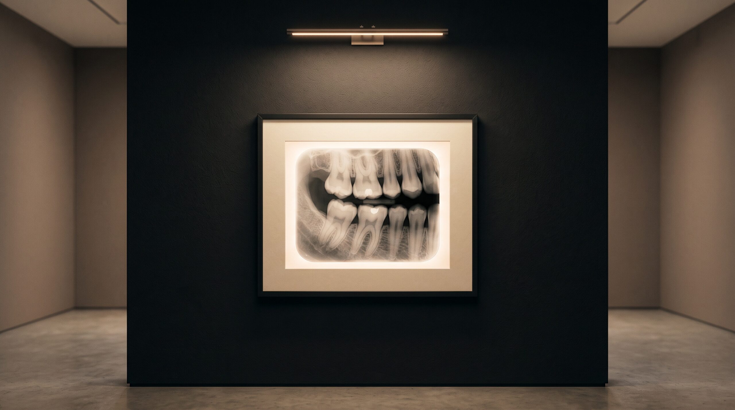
A dental bitewing radiograph presented as framed fine art on a gallery wall

01 Sep How AI Reads a Bitewing: Caries and Bone-Loss Detection, Explained

Posted at 09:44h in Dental Imaging by Raymond 0 Comments
0 Likes

A bitewing is a study in shadow, and shadow is exactly what the eye reads unreliably. Here is how a neural network learns to see the interproximal lesion and measure the bone crest - and why a confident overlay is only ever as good as...

Read More
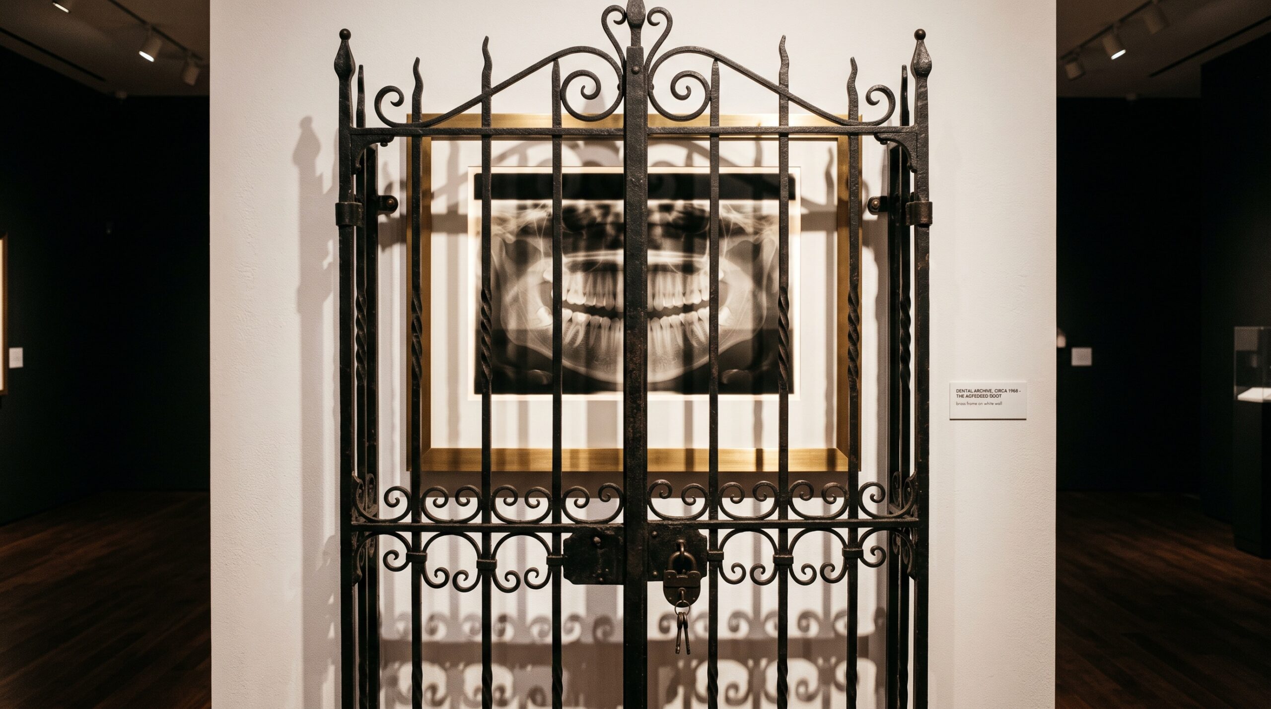
A framed dental radiograph glowing behind a closed, locked iron gate on a gallery wall - the image visible but unreachable.

31 Aug The Image That Cannot Travel: DICOM, Interoperability, and the Quiet Cost of a Radiograph That Will Not Move

Posted at 10:38h in Dental Imaging by Raymond 0 Comments
0 Likes

The finest radiograph in the world is worthless to the specialist who cannot open it cleanly. Between one practice and the next stands the quiet, unglamorous problem of interoperability - and the images that arrive flattened, stripped, or not at all....

Read More
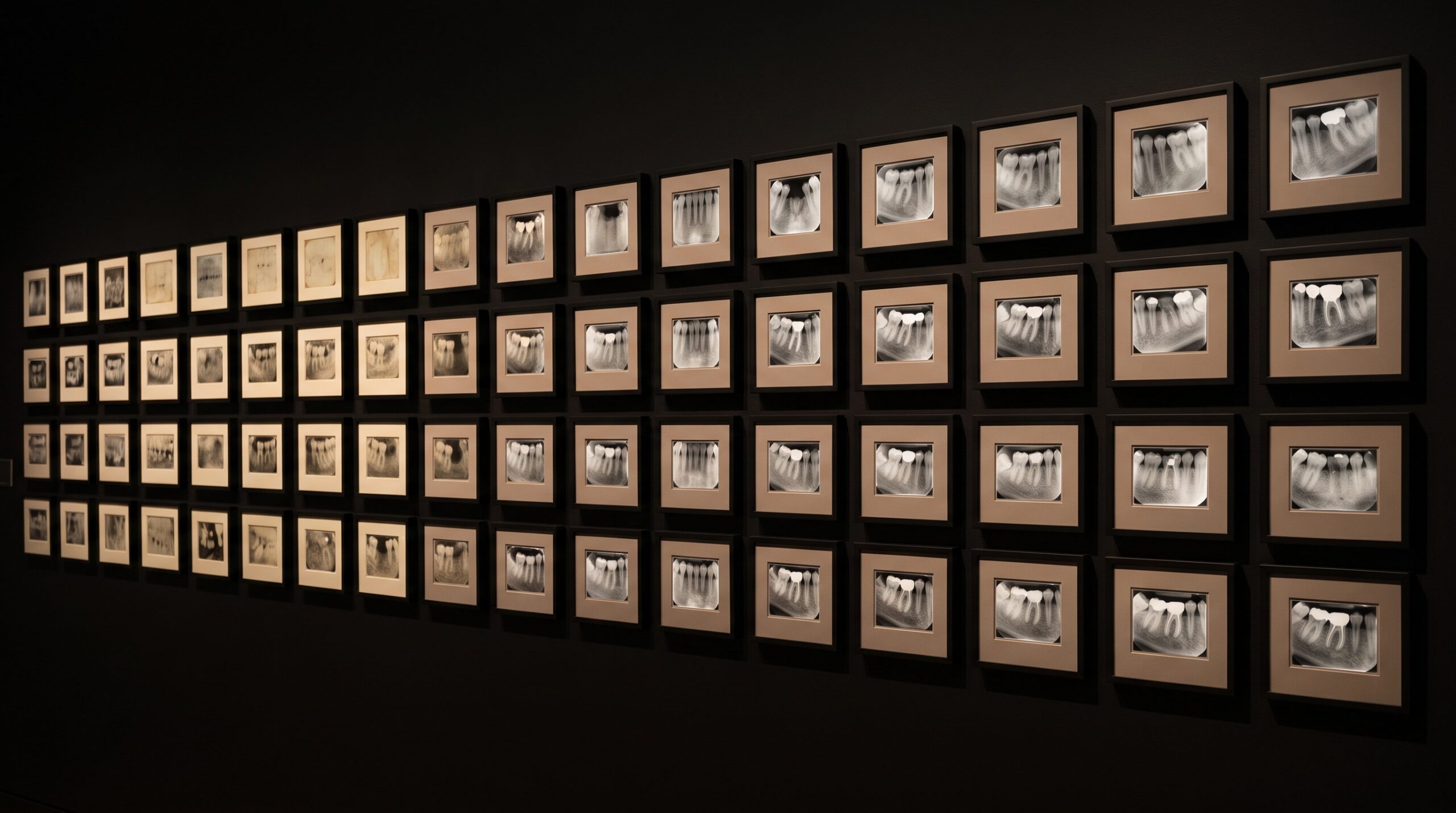
A gallery wall of small framed dental images arranged in a grid, progressing from faint and grainy to crisp and luminous

30 Aug Two Hundred Patents Deep: What a Two-Decade Portfolio Reveals About How the Dental Image Learned to See

Posted at 10:37h in Dental Imaging by Raymond 0 Comments
0 Likes

A single vendor recently tallied two decades of dental-imaging patents - hundreds of filings, a couple hundred granted. Read as more than a press release, it is a map of how the dental image was engineered, invention by patient invention....

Read More
A volumetric CBCT render of a jaw and teeth presented as suspended sculpture, its internal cross-section planes faintly visible

29 Aug Diagnocat on CBCT Volumes: 60+ 3D Findings and the API Path

Posted at 09:44h in Dental Imaging by Raymond 0 Comments
0 Likes

A CBCT scan is a quarter-million slices of latent information. Diagnocat turns that volume into a navigable 3D reading of 65+ conditions - but only after the DICOM leaves the operatory for the cloud. The findings are the art; the path is the plumbing....

Read More
  • 1
  • 2
  • 3
  • 4
  • 5
  • 6
  • 7
  • 8
  • 9
  • 10
  • 11
  • 12
  • 13
  • 14
© Copyright 2020, Raster Builders Inc. All Rights Reserved.